
Preparation method of carbon material sample for atomic force microscope contact mode characterization

An atomic force microscope, carbon material technology, applied in scanning probe microscopy, instruments, measurement devices, etc. question

Active Publication Date: 2019-12-13
SICHUAN UNIVERSITY OF SCIENCE AND ENGINEERING
View PDF10 Cites 0 Cited by
  • Summary
  • Abstract
  • Description
  • Claims
  • Application Information

AI Technical Summary

Problems solved by technology

[0009] However, the tapping mode cannot obtain the mechanical information of the sample surface, including the friction force of the sample surface, the force-distance curve between the sample and the needle tip
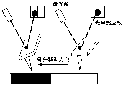
If the contact mode is used, the mechanical information can be obtained, but when there is a deficiency, when the sample and the base are not firm, the sample will be subjected to lateral force (friction force) during the measurement process, causing the sample to move laterally, so that it cannot be obtained. Clear images, and finally can not accurately obtain the mechanical information of the sample surface

Method used


Image

  • Preparation method of carbon material sample for atomic force microscope contact mode characterization
  • Preparation method of carbon material sample for atomic force microscope contact mode characterization
  • Preparation method of carbon material sample for atomic force microscope contact mode characterization

Examples


Embodiment 1

[0033] 1) Prepare 0.1 wt% epoxy resin organic polymer solution and let it stand for 24 hours.

[0034] 2) Dip the organic polymer solution with a glass rod to take a small amount of organic solution, coat the highly oriented graphite substrate once (only once), put it in a petri dish, dry naturally, and after removing the solvent, the surface is covered with a layer Flat polymer film.

[0035] 3) Evenly disperse graphene in deionized water, and disperse it ultrasonically for 30 minutes to obtain a dark black dispersion, and the concentration should not be too high.

[0036] 4) Use a dropper to take half a drop of dispersion liquid, evenly flow to the surface of the polymer film, and heat it with a hair dryer on the back of the highly oriented graphite substrate for 10s. Samples such as graphene can be fixed on the film, which can be used in AFM contact mode to stably measure its surface properties, and will not drift due to lateral force.

Embodiment 2

[0038] 1) Prepare 3 wt% polyvinyl alcohol organic polymer solution and let it stand for 24 hours.

[0039] 2) Dip the solution with a glass rod to take a small amount of polyvinyl alcohol organic solution, coat the base of the mica sheet once (only once), put it in a petri dish, let it dry naturally, and after removing the solvent, the surface is covered with a layer of surface The flat polymer film has a surface roughness of about 0.6 nm, such as Figure 4 , 5 As shown, it can meet the requirements of AFM testing.

[0040] 3) Evenly disperse graphene in anhydrous ethanol, disperse ultrasonically for 30 minutes, and obtain a dark black dispersion liquid, and the concentration should not be too high.

[0041] 4) Use a dropper to take half a drop of the dispersion, and evenly flow it onto the surface of the polymer film, and heat it with a hair dryer on the back of the mica sheet for 50s to fix the graphene on the film. The samples made in this way can be used for AFM Its sur...

Embodiment 3

[0043] 1) Prepare 5 wt% polyvinylidene fluoride organic polymer solution and let it stand for 24 hours.

[0044] 2) Dip the organic polymer solution with a glass rod to take a small amount of organic solution, coat the substrate of the silicon wafer once (only once), put it in a petri dish, dry it naturally, and after removing the solvent, the surface is covered with a layer of surface The flat polymer film has a surface roughness of about 0.6 nm.

[0045] 3) Disperse the carbon black evenly in acetone and disperse it ultrasonically for 30 minutes to obtain a dark black dispersion, and the concentration should not be too high.

[0046] 4) Use a dropper to take half a drop of dispersion liquid, evenly flow to the surface of the polymer film, and heat it with a hair dryer on the back of the silicon wafer substrate for 100s. The carbon black sample can be fixed on the film, which can be used in AFM contact mode to stably measure its surface properties, and it will not drift due ...


PUM


Abstract

The invention discloses a preparation method for a carbon material sample used for atomic force microscope contact mode representation. The method comprises the following steps of 1) preparing an organic macromolecule solution having a concentration of 0.1 to 5 wt% and allowing the solution to stand for 24h; 2) coating the organic high molecular solution onto a base material and drying the organic high molecular solution naturally to a constant weight to remove a solvent so that the surface of the base material is coated with a polymer film having a flat surface and a film-coated base material is obtained for application; 3) uniformly dispersing a carbon molecular material to be measured in a good solvent and subjecting the carbon molecular material to ultrasonic dispersion for 30 minutes to obtain a dark carbon molecular dispersion liquid for application; and 4) uniformly dropping the carbon molecular dispersion liquid on the side covered with the polymer film of the film base material, and then heating the other side of the film base material so that the carbon molecule material to be measured is fixed on the polymer film to obtain the carbon material sample. The prepared carbon material sample is to be subjected to stable determination of surface properties thereof in an AFM contact mode.

Description

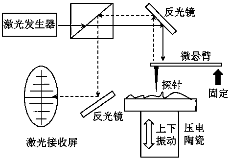
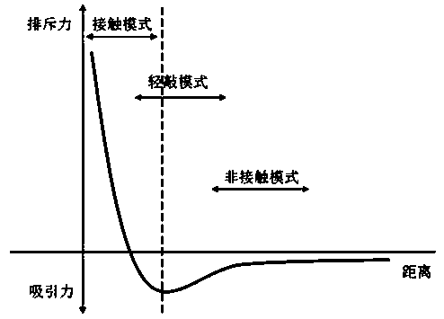
technical field [0001] The invention belongs to the technical field of nanomechanical measurement, and in particular relates to a method for preparing a carbon material sample for atomic force microscope contact mode characterization. Background technique [0002] The first AFM invented in 1986 made up for STM’s lack of sample conductivity. AFM uses a very sensitive micro-cantilever (force constant is 0.02-20N / m). The structure of AFM is as follows: figure 1 Shown: One end of the cantilever is fixed on the base, and the other end is fixed with a tapered tip with a tip curvature radius of nanometer (1~10nm). When the tip is close to the sample, there are attractive and repulsive forces between the sample surface and the tip The bending deformation of the cantilever will cause the position change of the laser reflection circuit on the photoelectric receiver, and the fluctuation response of the sample surface is the fluctuation of the force, so as to obtain the height image of ...

Claims


Application Information

Patent Timeline
Patent Type & Authority Patents(China)
IPC IPC(8): G01Q60/24
CPCG01Q60/24
Inventor 陈建龚勇代祖洋辜其隆林小力
Owner SICHUAN UNIVERSITY OF SCIENCE AND ENGINEERING
Learn More