Biological magnetic photoacoustic combined endoscopic imaging method

An imaging method and biomagnetic technology, applied in the field of medical imaging, can solve the problems of inability to observe and diagnose biological cavity tissue in a timely and effective manner, and achieve the effect of high spatial resolution

Inactive Publication Date: 2017-08-18
NORTH CHINA ELECTRIC POWER UNIV (BAODING)
View PDF5 Cites 7 Cited by
  • Summary
  • Abstract
  • Description
  • Claims
  • Application Information

AI Technical Summary

Problems solved by technology

Compared with endoscopic imaging, this method cannot timely and effectively observe and diagnos

Method used


Image

  • Biological magnetic photoacoustic combined endoscopic imaging method
  • Biological magnetic photoacoustic combined endoscopic imaging method
  • Biological magnetic photoacoustic combined endoscopic imaging method

Examples


Embodiment Construction

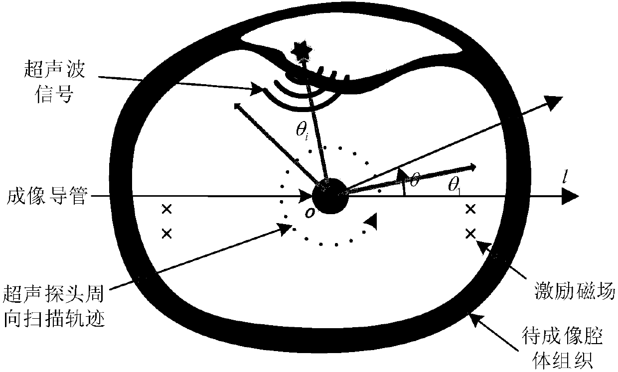
[0041] Endoscopic magneto-photo-acoustic (EMPA) imaging uses the same imaging system to simultaneously perform ultrasound, photoacoustic and inductive magnetoacoustic imaging inside the biological cavity. The ultrasonic transducer directly collects the ultrasonic signal reflected, scattered or generated by the tissue in the cavity, and then the combined image is obtained after fusion by the computer.

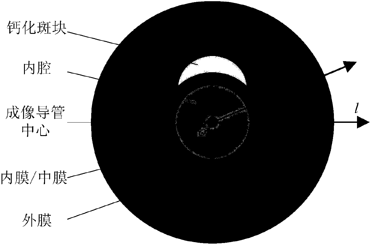
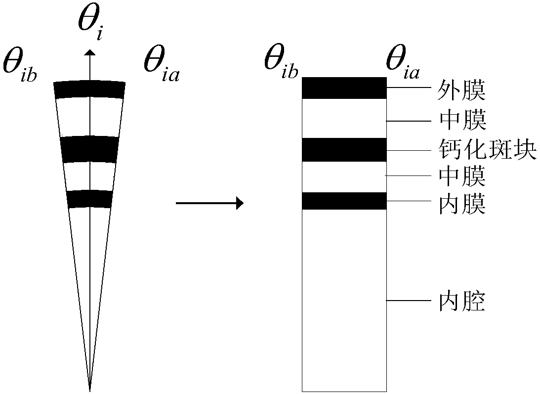
[0042] 1. Establish a cross-sectional model of multi-layer cavity tissue:

[0043] as attached figure 1 As shown, taking the cross-sectional model of a blood vessel as an example, the imaging catheter is located in the center of the model, and the ultrasonic transducer is located at the top of the imaging catheter. Surrounding the catheter from the inside to the outside along the radial direction are the vascular lumen, atherosclerotic plaque (calcification, lipid fibrous or mixed plaque), vessel wall intima / media and adventitia. The coordinate system where the model is locate...


PUM


Abstract

The invention relates to a biological magnetic photoacoustic combined endoscopic imaging method. The method comprises the following steps: on the basis of establishing a multi-layer cavity tissue cross section model, performing numerical simulation on the ultrasonic echo imaging, photoacoustic imaging and induction type magnetoacoustic imaging processes in a cavity channel to acquire a tissue reflected ultrasonic echo signal and tissue generated photoacoustic and magnetoacoustic signals on the cross section of the cavity channel; and performing optimal weighted summation on the three ultrasonic signals to acquired a fused combined imaging signal. The cavity tissue reflected/scattered ultrasonic echo signal and the tissue generated photoacoustic and magnetoacoustic signals which are received by an ultrasonic transducer in a time-share mode are fused in the signal layer. Compared with the traditional method, the method has the advantages that the combined imaging signal acquired by the method can retain the shape structure and the component information of the tissue more, the reestablished combined image has extremely high spatial resolution, contrast ratio, sensitivity and contrast resolution, and can accurately display the position, the shape and the functional components of each tissue in the wall of the cavity channel.

Description

technical field [0001] The invention relates to a method for performing magneto-optoacoustic combined endoscopic imaging on biological cavity tissue, and belongs to the technical field of medical imaging. Background technique [0002] Biophotoacoustic (PA) imaging is based on the photoacoustic effect of biological tissue as the physical basis, that is, the tissue absorbs short-pulse laser light and then heats up and expands to generate ultrasonic waves (ie, photoacoustic signals). Inductive magnetoacoustic tomography (magnetoacoustictomography with magnetic induction, MAT-MI) imaging is based on the magnetoacoustic effect of biological tissues, that is, the target is placed in a static magnetic field, and magnetic pulse excitation is applied in the same direction as the static magnetic field. The action of the eddy current induced in the medium and the static magnetic field produces the Lorentz force, and the charged particles vibrate under the action of the Lorentz force, t...

Claims


Application Information

Patent Timeline
IPC IPC(8): A61B5/00
CPCA61B5/0093A61B5/0095
Inventor 孙正杨凯旋
Owner NORTH CHINA ELECTRIC POWER UNIV (BAODING)
Why Eureka
Try Eureka