
Method for detecting circulating tumor cell in whole blood by utilizing ICP-MS and fluorescence imaging

An ICP-MS, fluorescence imaging technology, applied in the field of biological analysis, can solve the problems of magnetic particle loss, sensitivity reduction, signal fluctuation, etc., to eliminate errors, improve stability and reliability, strong simultaneous analysis and accurate quantification.

Inactive Publication Date: 2017-08-18
WUHAN UNIV
View PDF0 Cites 3 Cited by
  • Summary
  • Abstract
  • Description
  • Claims
  • Application Information

AI Technical Summary

Problems solved by technology

However, magnetic separation is inherently flawed in that the magnetic particles are partially lost during the washing step, which causes measurement errors, signal fluctuations, and reduced sensitivity

Method used


Image

  • Method for detecting circulating tumor cell in whole blood by utilizing ICP-MS and fluorescence imaging
  • Method for detecting circulating tumor cell in whole blood by utilizing ICP-MS and fluorescence imaging
  • Method for detecting circulating tumor cell in whole blood by utilizing ICP-MS and fluorescence imaging

Examples


Embodiment l

[0036] Example 1: Preparation of Amino-modified Cs-doped Multinuclear Magnetic Nanoparticles, MMNPs-anti-EpCAM Probe Magnetic Balls and QDs-anti-ASGPR Labeled Probes

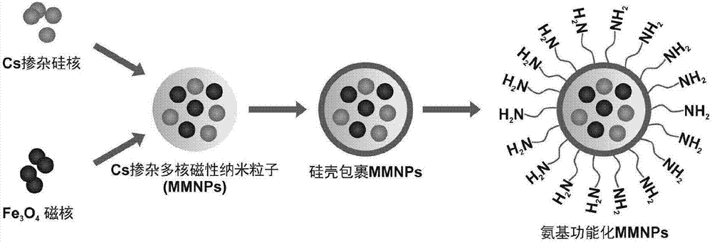
[0037] Schematic diagram of the preparation process of amino-modified Cs-doped multinuclear magnetic nanoparticles. figure 1 As shown, the specific steps are as follows:

[0038] (1) Preparation of magnetic nanoparticles Fe by co-precipitation method 3 o 4 : First weigh 1.35g FeCl 3 ·6H 2 O, 0.5gFeCl 2 4H 2 O, add 25mL deionized water; after the solid is completely dissolved, mechanically stir in an argon atmosphere and heat to reflux at 80°C; when the color of the solution turns dark orange, increase the argon flow rate, and quickly add 12.5mL concentrated ammonia water , at this time, the color of the solution changed from orange to black; continue heating and stirring for 20min, and then cool to room temperature to obtain magnetic nanoparticles Fe3 o 4 , using the magnetic separation method to wash the...

Embodiment 2

[0043] Example 2: Characterization of amino-modified Cs-doped multinuclear magnetic nanoparticles

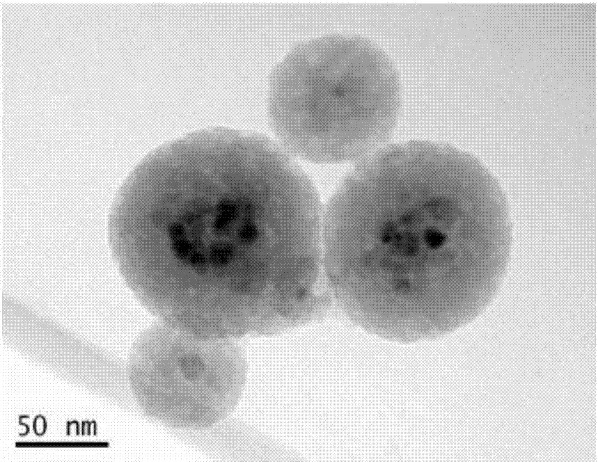
[0044] Adopt transmission electron microscope (TEM) to observe the MMNPs-NH that embodiment 1 prepares 2 , its TEM picture is as figure 2 shown. from figure 2 It can be seen that MMNPs-NH 2 Good dispersion, relatively uniform particle size, about 100-150nm, it can be clearly observed that a magnetic ball contains multiple Fe 3 o 4 nuclear.

Embodiment 3

[0045] Example 3: ICP-MS analysis of target cells and non-target cells

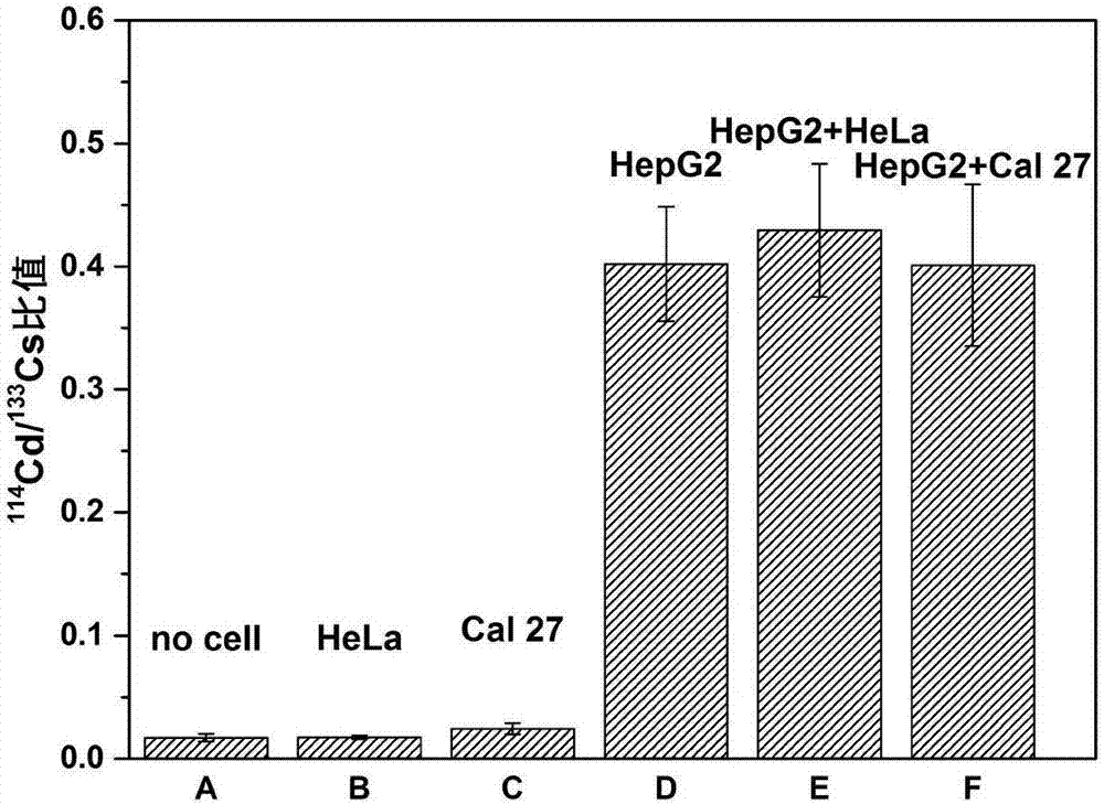
[0046] In this example, cadmium selenide (CdSe) quantum dots were used as labeled quantum dots, HepG2 cells expressing ASGPR antigen (liver-specific transmembrane glycoprotein) were selected as target objects, and human tongue squamous cell carcinoma cells Cal 27 that did not express ASGPR were selected. cells and human cervical cancer HeLa cells as negative control cells. Group A contains HepG2 cells, group B contains 1×10 4 HeLa cells, group C contains 1×10 4 Cal-27 cells, group D contains 1×10 4 HepG2 cells, group E contained 1×10 4 HepG2 cells and 1×10 5 HeLa cells, group F contained 1×10 4 HepG2 cells and 1×10 5 Cal-27 cells.

[0047] Take 2 μL prepared MMNPs-anti-EpCAM probe magnetic balls, block with 200 μL bovine serum albumin solution (1% BSA) for 0.5 h, in order to reduce the non-specific adsorption of the probe; then add the cells to be tested and place on a shaker at 90 rpm Incubate at...


PUM

PropertyMeasurementUnit
particle diameteraaaaaaaaaa

Abstract

The invention discloses a method for detecting a circulating tumor cell (CTC) in whole blood by utilizing ICP-MS and fluorescence imaging. The method comprises the steps of capturing the circulating tumor cell in a whole blood sample for carrying out magnetic separation through antibody-coated Cs-doped multinuclear magnetic nanoparticles; then adding antibodies modified by quantum dots to carry out fluorescence labeling, adopting the quantum dots as common labels for ICP-MS and fluorescence imaging, using a fluorescence microscope for imaging the CTC or directly introducing ICP-MS detection after dispersing the CTC through nitric acid, and realizing quantitative counting of the CTC through elements in the quantum dots labeled on the ICP-MS detection antibodies. The method provided by the invention has the advantages of high selectivity, high sensitivity, matrix interference resistance and the like; Cs doped in the magnetic nanoparticles can be used as an internal standard element, and an internal standard method determination is adopted so as to eliminate the error caused on experimental result determination by the loss of the magnetic nanoparticles during an analysis process, so that the reliability of the method is further improved. The method provided by the invention is applicable to human whole blood sample analysis, and has a favorable application prospect clinically.

Description

technical field [0001] The invention belongs to the field of biological analysis methods, in particular to a method for detecting circulating tumor cells (CTCs) in whole blood by using ICP-MS and fluorescence imaging. Background technique [0002] According to data released by the National Cancer Center of my country, in 2015, there were 4.292 million new cancer cases and 2.814 million cancer deaths in my country. The incidence of liver cancer ranks fourth after lung cancer, gastric cancer, and esophageal cancer, while the mortality rate of liver cancer is second only to lung cancer and gastric cancer. At the same time, liver cancer is also one of the most common cancers in the world, and there is no effective cure, especially in the advanced stage of liver cancer. Most cancer-related deaths are due to metastases from the original cancer cells. Like many other diseases, early diagnosis is critical for effective treatment of cancer. The development of a feasible protocol f...

Claims


Application Information

Patent Timeline
Patent Type & Authority Applications(China)
IPC IPC(8): G01N33/574G01N33/543G01N33/533G01N27/62G01N21/64
CPCG01N21/6402G01N21/6428G01N21/6452G01N21/6458G01N27/626G01N33/533G01N33/54326G01N33/574G01N33/57438G01N2021/6441G01N2446/90G01N2800/085
Inventor 胡斌杨彬何蔓陈贝贝
Owner WUHAN UNIV
Learn More