Application of IL-12p40(-/-)IL-2R alpha(-/-) mouse model
1. The technology of il-12p40 and mouse model, applied in the field of medical biology, can solve the problems of lacking animal models of autoimmune myelofibrosis
- Summary
- Abstract
- Description
- Claims
- Application Information
AI Technical Summary
Problems solved by technology
Method used
Image
Examples
Embodiment 1
[0033] Example 1. Establishment of IL-12p40(- / -)IL-2Rα(- / -) mice
[0034] IL-2Rα (- / -) mice (B6.129S4-Il2ratm1Dw) [Willerford, D.M., et al. (1995). Immunity 3 (4): 521-530] of C57BL / 6J background and IL-12p40 (- / -) mouse (B6.129S1-Il12btm1Jm) [Magram, J., et al. (1996). Immunity 4 (5): 471-481] was purchased from Jackson Laboratory, USA (The Jackson Laboratory, Maine, USA, http : / / www.jax.org / ), reared in a special pathogen-free (SPF) environment.
[0035] We crossed IL-2Rα(+ / -) mice with IL-12p40(- / -) mice to obtain IL-12p40(+ / -)IL-2Rα(+ / -) mice; 12p40(- / -) mice were crossed to obtain IL-12p40(- / -)IL-2Rα(+ / -) mice. The IL-12p40(- / -)IL-2Rα(- / -) mice and IL-12p40(- / -)IL-2Rα(+ / -) mice used in the experiment were all produced by IL-12p40(- / -) IL-2Rα(+ / -) mice were bred. Since the mutant genes of IL-12p40 and IL-2Rα both contain a neo gene, the method for identifying the mouse IL-12p40 gene is to use genotyping to identify the wild-type gene of IL-12p40 and identify the mous...
Embodiment 2
[0036] Embodiment 2. routine blood test
[0037] The peripheral blood of the mice was collected into an anticoagulant tube, 150 μL of blood was drawn, and blood routine data were collected by an automatic hematology analyzer.
[0038] see results figure 1 , Compared with IL-12p40(- / -)IL-2Rα(+ / -) mice, the peripheral red blood cell count (RBC) of IL-12p40(- / -)IL-2Rα(- / -) mice, Hemoglobin content (HGB), hematocrit (HCT) and white blood cell count (WBC) all decreased significantly, but platelet count (PLT) did not change significantly.
Embodiment 3
[0039] Example 3. Histopathological examination
[0040] The liver, spleen and bone marrow tissues of the mice were fixed in 4% neutral formaldehyde for 1-2 days. The bone marrow tissue was put into decalcification solution (5% hydrochloric acid+5% acetic acid) for 24 hours. Tissues were dehydrated and embedded in paraffin, and sectioned into 4 μm slices. H&E staining was performed after dewaxing the thin sections. Bone marrow tissue showed fibrosis by silver staining of reticular fibers.
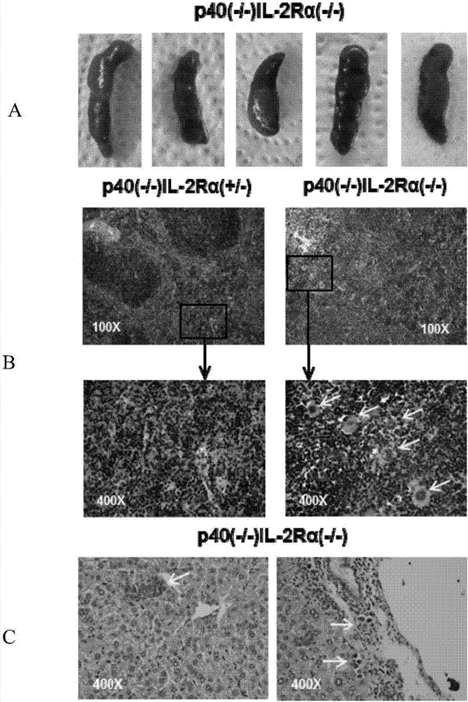
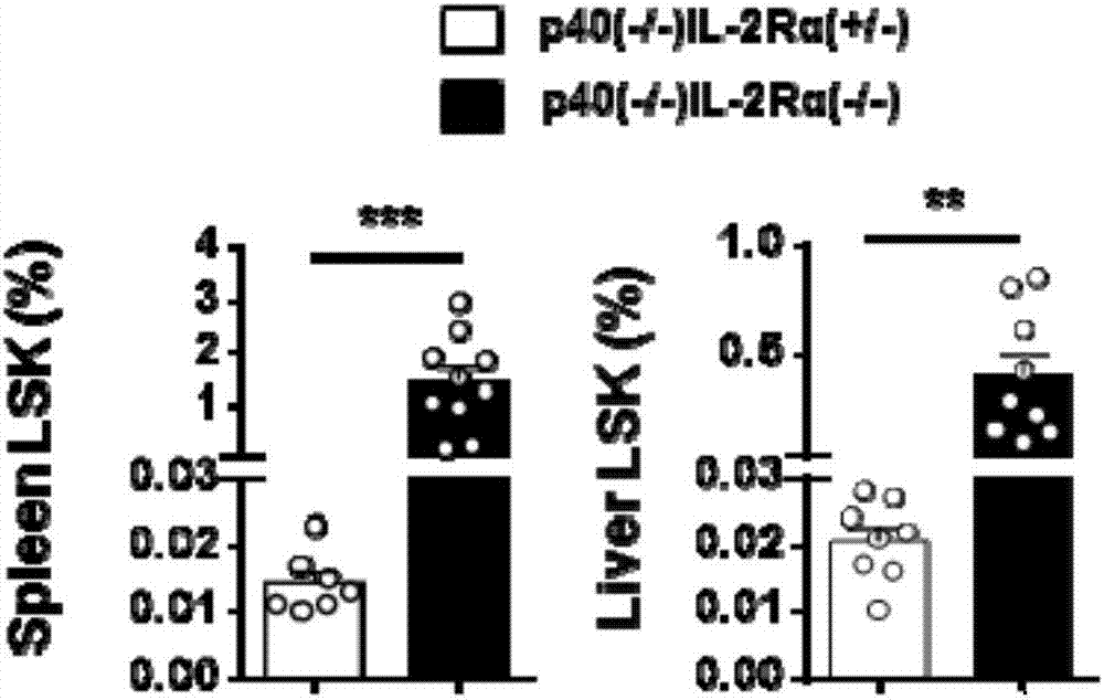
[0041] see results figure 2 , image 3 ,From figure 2 Spleen of IL-12p40(- / -)IL-2Rα(- / -) mice compared with IL-12p40(- / -)IL-2Rα(+ / -) mice can be seen in HE staining Structural changes occurred, the distinction between red and white pulp was not obvious, and a large number of megakaryocytes appeared. Hematopoietic islands exist in the liver of IL-12p40(- / -)IL-2Rα(- / -) mice. These results demonstrate extramedullary hematopoiesis in the spleen and liver of IL-12p40(- / -)IL-2Rα(- / -) mi...
PUM
 Abstract
Description
Claims
Application Information
 - R&D
- Intellectual Property
- Life Sciences
- Materials
- Tech Scout
- Unparalleled Data Quality
- Higher Quality Content
- 60% Fewer Hallucinations
Browse by: Latest US Patents, China's latest patents, Technical Efficacy Thesaurus, Application Domain, Technology Topic, Popular Technical Reports.
© 2025 PatSnap. All rights reserved.Legal|Privacy policy|Modern Slavery Act Transparency Statement|Sitemap|About US| Contact US: help@patsnap.com


