Method and device for preparing in-vitro three-dimensional tissue model

A technology for preparing devices and tissues, applied in the field of preparation of in vitro three-dimensional tissue models, can solve problems such as difficulty in manipulating the printing speed of cells, and achieve the effects of contributing to accuracy and rapid prototyping, strong applicability, and high survival rate

Active Publication Date: 2017-11-07
SHENZHEN GRADUATE SCHOOL TSINGHUA UNIV
View PDF11 Cites 14 Cited by
  • Summary
  • Abstract
  • Description
  • Claims
  • Application Information

AI Technical Summary

Problems solved by technology

[0006] The main purpose of the present invention is to overcome the deficiencies of the prior art and provide a method and device for preparin

Method used


Image

  • Method and device for preparing in-vitro three-dimensional tissue model
  • Method and device for preparing in-vitro three-dimensional tissue model
  • Method and device for preparing in-vitro three-dimensional tissue model

Examples


preparation example Construction

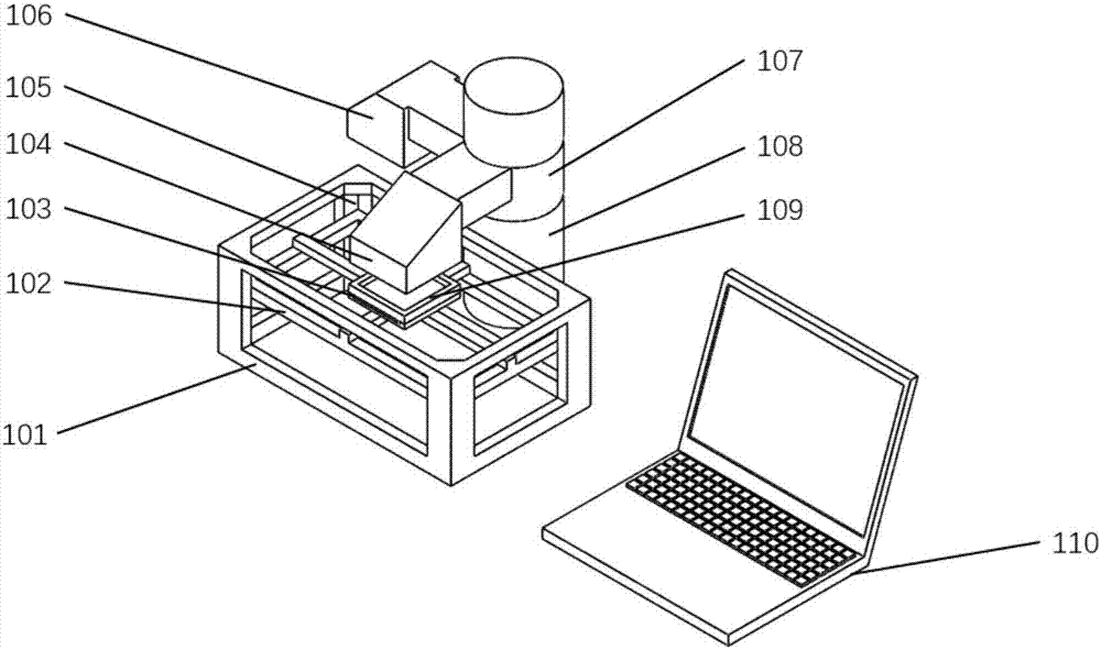
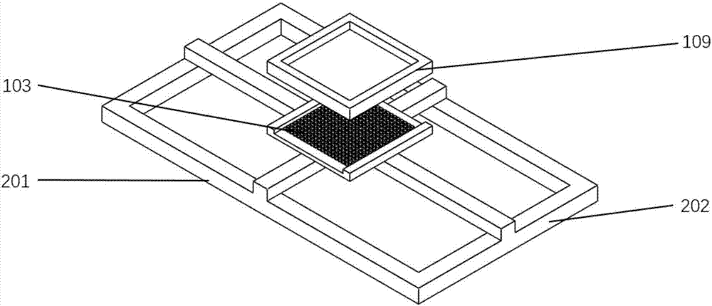
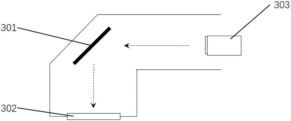
[0043] refer to Figure 4 to Figure 7 , in another embodiment, a method for preparing an in vitro three-dimensional tissue model, comprising the following steps:

[0044] (1) Using computer-aided design software to design a three-dimensional tissue model in vitro, layering the designed tissue model one by one, obtaining each layer of tissue after layering, and generating tissue pattern data of each layer. The thickness of each layer can be selected in the range of 100μm-1mm according to the needs, and the different thickness of each layer can be realized by pouring different doses of the compound solution.

[0045] (2) Mix the cells, growth factors and collagen solution evenly to obtain a mixed solution, then inject photocurable hydrogel into the mixed solution, and then add a photoinitiator to obtain a photocurable composite solution mixed with cells. According to the needs of tissue construction, different cells can be used in different layers of the tissue, and the growth ...

Embodiment 1

[0050] The preparation of the three-dimensional liver lobular tissue model in vitro, the specific steps are as follows:

[0051] (1) According to the characteristics of the hepatic lobular tissue structure, a three-dimensional hepatic lobular tissue model in vitro was designed by using computer-aided design software, and the designed tissue model was layered one by one. After layering, each layer of tissue was obtained, and the tissue pattern data of each layer was generated. The thickness of each layer is 100 μm.

[0052] (2) Mix the cells, growth factors and collagen solution evenly to obtain a mixed solution, then inject photocurable hydrogel into the mixed solution, and then add a photoinitiator to obtain a photocurable composite solution mixed with cells. Wherein the cells are human liver cells (702), endothelial cells and mesenchymal cells (701), the collagen in the collagen solution is type I collagen, the mixing density of the cells in the mixed solution is 1×〖10〗7 cel...


PUM


Abstract

The invention provides a method and a device for preparing an in-vitro three-dimensional tissue model. The method comprises the following steps: (2) preparing a photocuring composite solution mixed with cells from an extracellular matrix material containing cells and growth factors, photocuring hydrogel and a photoinitiator; (3) adding the prepared photocuring composite solution to a forming tray, generating a changing controllable electric field by a control electrode array, and manipulating the cells by the aid of dielectrophoretic force to enable the cells to move and reach a target area; (4) importing a layer of tissue pattern data into a DMD (digital micromirror device), generating a photomask of the layer of tissue pattern, and curing the photocuring composite solution with a surface exposure technology; (5) after photocuring of the layer of the tissue is finished, repeating steps (3) and (4) for cell manipulation and photocuring for the next layer of tissue, and performing accumulation layer by layer to obtain the in-vitro three-dimensional bionic tissue model. Problems that the cells are difficult to manipulate, printing speed is low and the like in the prior art can be solved.

Description

technical field [0001] The invention relates to the technical field of in vitro tissue model preparation, in particular to a preparation method and device for an in vitro three-dimensional tissue model. Background technique [0002] Tissue and organ defects or dysfunction caused by various reasons are the main causes of harm to human health. The repair and functional reconstruction of tissue and organ defects are challenges in the medical field. The development of tissue engineering has brought new hope for the treatment of tissue and organ damage and loss of function. Applying engineering and biological principles, using biomaterials, cells and growth factors, etc. in combination or alone, reconstitutes tissue and organ models in vitro. Simulating factors such as tissue development, local microenvironment and biomechanical stimuli is of great help to the research of pathology and pharmacology. [0003] In recent years, 3D bioprinting technology has become more and more wid...

Claims


Application Information

Patent Timeline
IPC IPC(8): A61L27/40A61L27/36A61L27/24A61L27/20A61L27/18A61L27/54A61L27/50A61L27/52B29C64/129B33Y10/00
CPCA61L27/18A61L27/20A61L27/24A61L27/36A61L27/3633A61L27/50A61L27/52A61L27/54B33Y10/00
Inventor 弥胜利孙伟蒲海涛徐圆圆
Owner SHENZHEN GRADUATE SCHOOL TSINGHUA UNIV
Why Eureka
Try Eureka