A cell imaging method

A cell and imaging technology, applied in the direction of material excitation analysis, fluorescence/phosphorescence, instruments, etc., can solve the problems of weakening fluorescence intensity, occupying excitation and emission channels, time-consuming and labor-intensive, etc., to reduce the influence of cells and increase selectivity , The effect of simplifying the experimental operation

Active Publication Date: 2020-06-30
PEKING UNIV
View PDF6 Cites 0 Cited by
  • Summary
  • Abstract
  • Description
  • Claims
  • Application Information

AI Technical Summary

Problems solved by technology

[0003] At present, the main application of LSCM is to collect the fluorescent signal of the sample for imaging, and to track the changes of cells in three-dimensional space. However, if the cells do not have fluorescence, they will be dark in the field of view of the fluorescence microscope, and it is difficult to locate the cells, which makes this method in practice. Encountered the problem of three-dimensional positioning of cells in:
Disadvantages: First, the operation of fluorescent labeling is relatively complicated, requiring exploration of conditions, time-consuming and laborious, and high cost; second, after the cells are labeled with fluorescent substances, they will occupy certain excitation and emission channels, which restricts the selection range of other fluorescent labeling substances in the experiment ; Third, there is a problem of photobleaching: since the fluorescence intensity of most fluorescent substances will be significantly weakened after long-term and intense laser irradiation, they cannot be detected under LSCM for a long time, so it is inevitable in imaging and quantification. System errors are generated; fourth, in the continuous observation of the dynamic process of drug uptake by cells, before the real-time observation of the fluorescently labeled drug enters the cells, the cells that are not fluorescently labeled cannot be accurately focused; when the cells are fluorescently labeled, the fluorescent The probe will have a certain effect on the state of the cell, and experimental errors will inevitably occur

Method used


Image

  • A cell imaging method
  • A cell imaging method
  • A cell imaging method

Examples


Embodiment 1

[0067] Example 1: LSCM detection and spectral scanning of unlabeled cells.

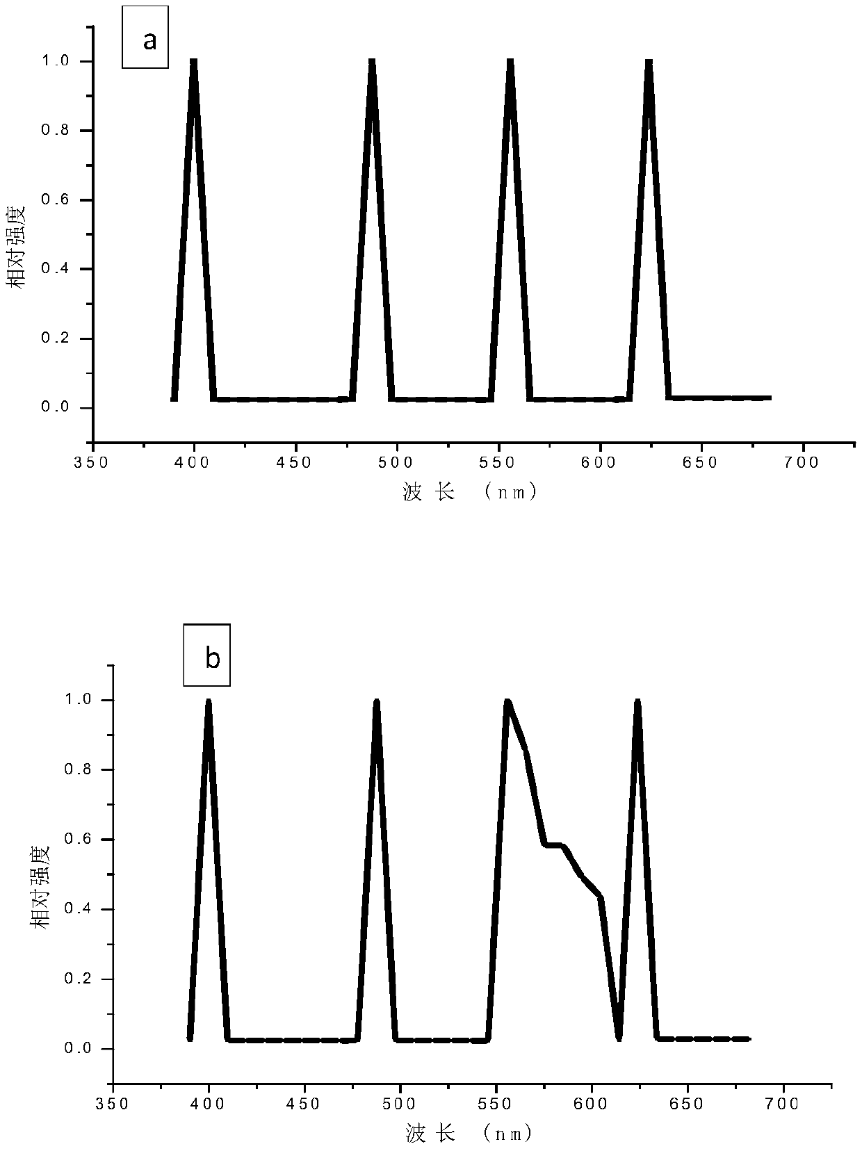
[0068] A549 cells were planted in a confocal glass dish, and the cells were adhered to the appropriate density and placed under an LSCM microscope. In the process of spectral scanning, four sets of excitation light source channels of LSCM are turned on, and lasers of four wavelengths are emitted: UV-405nm, Ar-488nm, Kr-561nm, HeNe-633nm, the detection wavelength range is set to 400nm to 680nm, and the step distance is 5nm. Plot the intensity-wavelength curve. Test results such as figure 1 Shown in (a) is the emission spectrum of A549 cells in the range of receiving wavelengths. There is a sharp peak at 405, 488, 561, and 633nm on the spectrum of A549 cells, and the peak width is only about 10nm. These peaks are all at the position of the excitation wavelength.

Embodiment 2

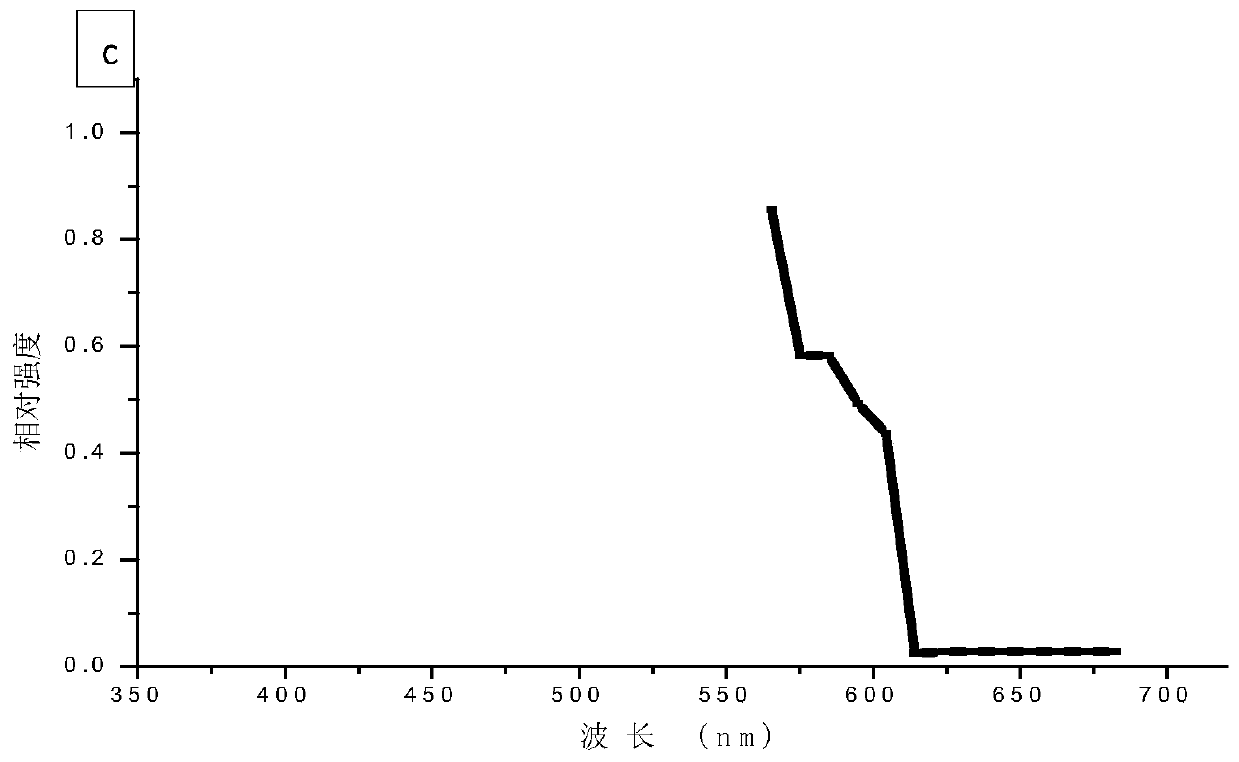
[0069] Example 2: LSCM detection and spectral scanning of cells labeled with DiI fluorescence.

[0070] A549 cells were planted in a confocal glass dish, and the cells were adhered to the wall and cultured to a suitable density. The cell membrane was fluorescently labeled with DiI, and then placed under an LSCM microscope for detection. DiI solution was used as the control group. In the process of spectral scanning, four sets of excitation light source channels of LSCM are turned on, and lasers of four wavelengths are emitted: UV-405nm, Ar-488nm, Kr-561nm, HeNe-633nm, and the detection wavelength range is set to 400nm to 680nm, and the step distance is 5nm. Plot the intensity-wavelength curve. Test results such as figure 1 Shown in (b) and (c) are the emission spectrum diagrams of the DiI fluorescently labeled cells and the DiI solution in the receiving wavelength range of 400-680 nm, respectively. Where (b) represents DiI fluorescently labeled cells, and (c) represents DiI...

Embodiment 3

[0072] Example 3: Cells labeled with DiI and Hoechst3332 fluorescence are detected by LSCM

[0073] A549 cells were planted in a confocal glass dish. After the cells adhered to the wall and cultured to a suitable density, the cell membrane was fluorescently labeled with DiI, and the cell nucleus was labeled with Hoechst3332, and then placed under an LSCM microscope for detection.

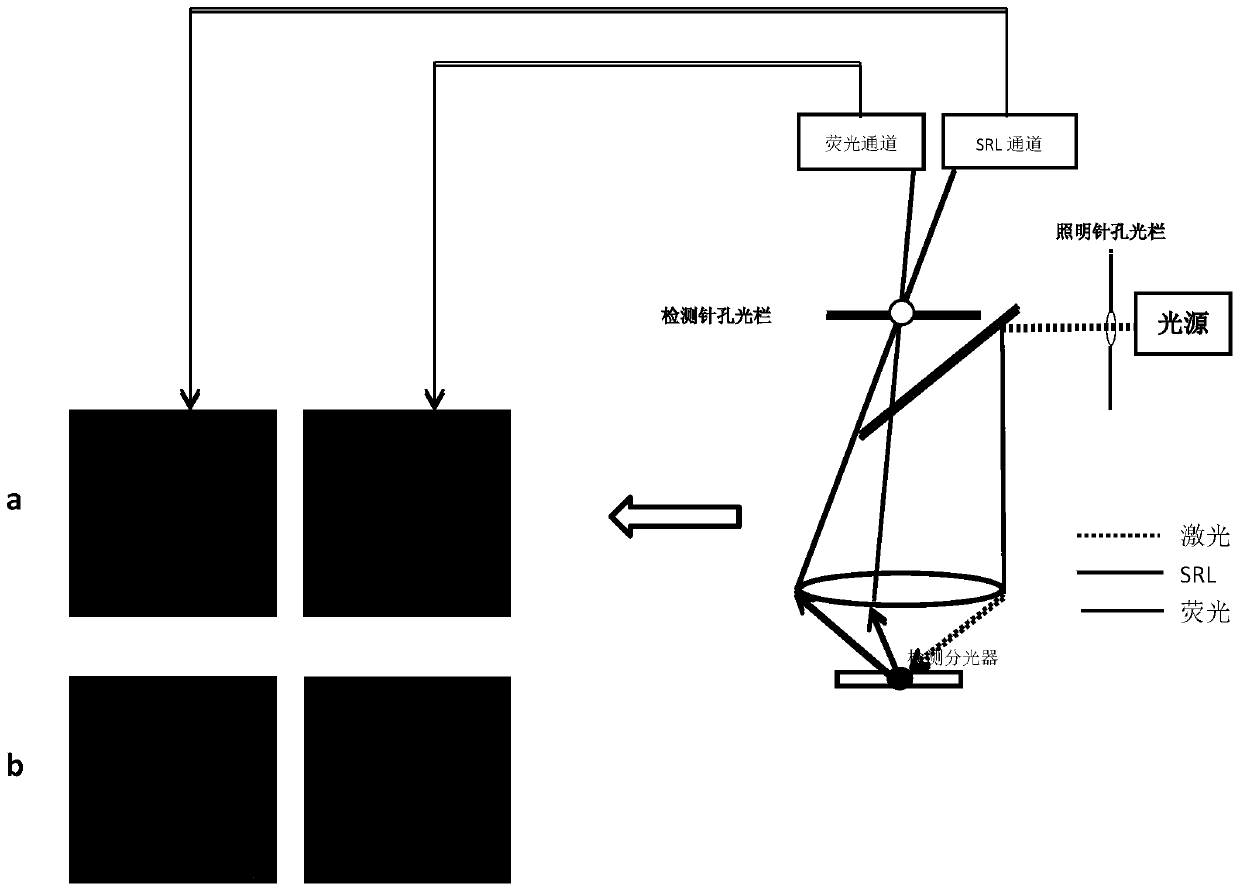
[0074] image 3 For cells labeled with DiI and Hoechst33342 fluorescence, LSCM was used for planar imaging, where: (b) is the partially enlarged image of (a), and (c) is the xzy longitudinal section image of (b). As shown in the figure, the SRL channel can clearly image the cells and has co-localization with DiI.

[0075] Figure 4 It is the image of the cell samples of each layer scanned continuously along the z-axis under LSCM, in which: (a) Hoechst33342 fluorescence channel, (b) DiI fluorescence channel, (c) SRL channel, (d) overlay of a, b, c. As shown in the figure, both the SRL channel and ...


PUM

PropertyMeasurementUnit
wavelengthaaaaaaaaaa

Abstract

The invention relates to a cell and tissue imaging method which comprises the following steps: 1) preparing a biological cell or tissue sample, wherein the sample can be live or dead (such as immobilized) cells or tissue; 2) performing Scattered and reflected light, SRL (scattered and reflected light) image acquisition, processing and analysis on the cell and tissue sample by using functions of various microscopes (by taking a laser scanning confocal microscope as an example).

Description

technical field [0001] The present invention relates to a cell and tissue imaging method, and in particular to the acquisition of SRL images alone and in combination with fluorescent or transmitted light images of cells and tissue samples using LSCM. Background technique [0002] Cells are the basic structural and functional units of organisms. It is known that all organisms except viruses are composed of cells, but the life activities of viruses must also be reflected in cells. Today, almost all basic research and key difficulties in life science research are closely related to cells. Taking cells as the research object and studying the various changes that occur in it, a very important link is imaging observation. At present, the methods for cell imaging mainly include: optical microscope technology includes ordinary compound optical microscope, fluorescence microscope, laser scanning microscope (including single-photon or multi-photon laser scanning microscope, super-res...

Claims


Application Information

Patent Timeline
Patent Type & Authority Patents(China)
IPC IPC(8): G01N21/64G01N21/39
CPCG01N21/39G01N21/64G01N21/6402G01N21/6428G01N21/6458G01N2021/6419G01N2021/6421G01N2021/6423G01N2021/6439
Inventor 袁兰徐萍梁磊杨凌飞李昀倩
Owner PEKING UNIV
Why Eureka
Try Eureka