Three-dimensional tumor model decellularization porous scaffold, construction method and application thereof

A technology of porous scaffolds and construction methods, applied in the direction of tumor/cancer cells, cell culture support/coating, animal cells, etc., can solve the problems of lack of flexibility in operation, large demand for decellularization reagents, etc., to facilitate cell adhesion , The decellularization process is simple and easy, and the effect of low immunogenicity

Active Publication Date: 2018-05-04
DALIAN UNIV OF TECH
View PDF8 Cites 10 Cited by
  • Summary
  • Abstract
  • Description
  • Claims
  • Application Information

AI Technical Summary

Problems solved by technology

This method often requires a peristaltic pump to provide pressure to introduce the decellularization reagent, and the demand for the decellularization reagent is large
In addition, the current decellularized lung scaffolds are mainly used to replace damaged lungs. The decellularization is aimed at the entire lung tissue, and the process of decellularization and reinfusion of cells needs to be completely sterile, and the operation lacks flexibility.

Method used


Image

  • Three-dimensional tumor model decellularization porous scaffold, construction method and application thereof
  • Three-dimensional tumor model decellularization porous scaffold, construction method and application thereof
  • Three-dimensional tumor model decellularization porous scaffold, construction method and application thereof

Examples


Embodiment 1

[0033] Example 1 Preparation of acellular pig lung three-dimensional tumor model scaffold

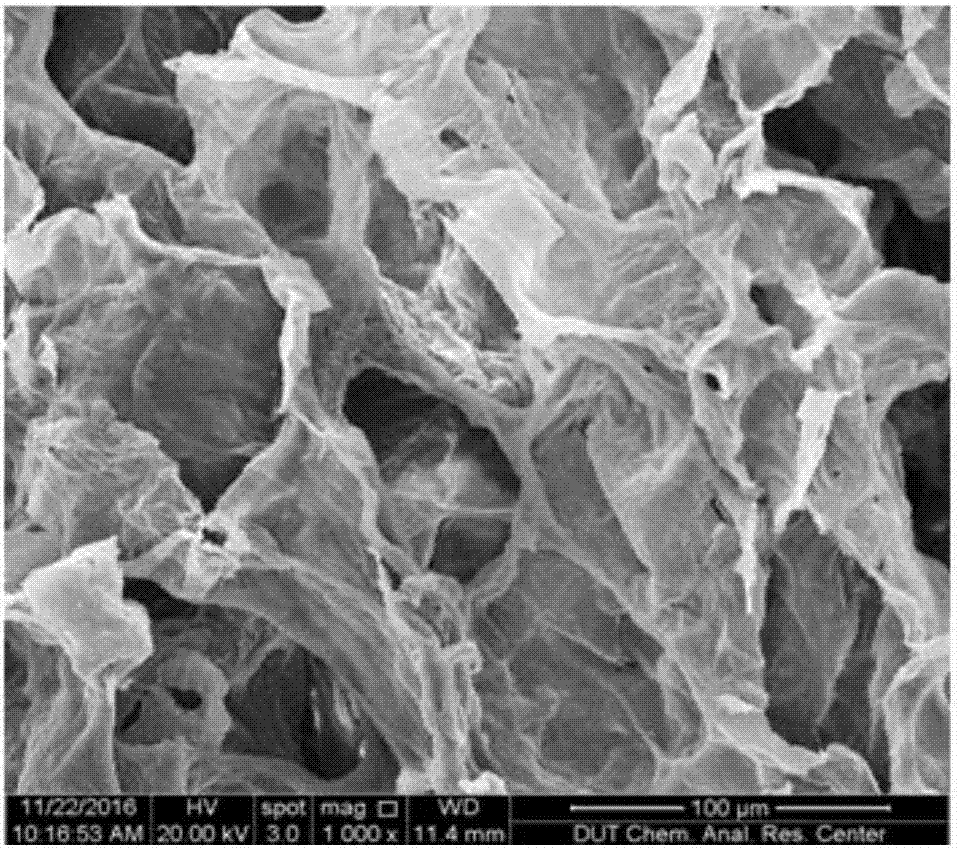
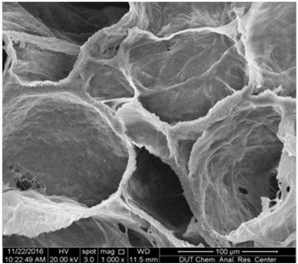
[0034] Place the whole pig lung tissue in the refrigerator at -20℃ for 6 hours. After it is fixed and formed, cut and observe the uniform part of the large bronchus with naked eyes. The size of the cut is 8cm. 3 . Put the pork lung cut into a beaker, then add 2000mL of double distilled water, magnetically stir for 30 minutes and then replace with new distilled water. After the above operation is repeated 3 times, wash twice with 2000mL, pH=7.4 PBS, each time with magnetic stirring 30min. After there is no obvious blood color in the tissue, add 2000 mL of 1% (wt%, the same below) SDS solution, and after magnetic stirring for 6 hours, replace with a new 1% SDS solution and continue stirring for 6 hours to remove cells. Then add 2000 mL of 0.5% (v%, the same below) TritonX-100 solution and stir for 10 hours to completely remove the cells. Subsequently, 2000mL PBS was added to wash 3 times, ...

Embodiment 2

[0035] Example 2 Preparation of acellular pig lung three-dimensional tumor model scaffold

[0036] Place the whole pig lung tissue in a refrigerator at -30°C for 3 hours. After it is fixed and formed, cut and observe the uniform parts of the large bronchus with naked eyes. The size of the cut is 6cm. 3 . Cut the pig lung into a beaker, then add 1000 mL of double-distilled water, magnetically stir for 40 minutes, and replace with new distilled water. After the above operations 5 times, wash the same 3 times with 1500 mL, pH=7.4 PBS, each for 30 minutes. After there is no obvious blood color in the tissue, add 1500 mL of 0.5% SDS solution, and after magnetic stirring for 6 hours, replace with a new 0.5% SDS solution and continue stirring for 12 hours to remove cells. Then add 2000mL of 0.3% TritonX-100 solution and stir for 12h to completely remove the cells. Subsequently, 1500mL PBS was added to wash 3 times, each duration of 1h, to completely wash the decellularization reagent. ...

Embodiment example 3

[0037] Implementation case 3 Cross-linking and performance test of acellular pig lung three-dimensional tumor model scaffold

[0038] Place the whole pig lung tissue in the refrigerator at -20℃ for 6 hours. After it is fixed and formed, cut and observe the uniform part of the large bronchus with naked eyes. The size of the cut is 8cm. 3 . Put the pork lung cut into a beaker, then add 2000mL of double distilled water, magnetically stir for 30 minutes and then replace with new distilled water. After the above operation is repeated 3 times, wash twice with 2000mL, pH=7.4 PBS, each time with magnetic stirring 30min. When there is no obvious blood color in the tissue, add 2000 mL of 1% SDS solution, and after magnetic stirring for 6 hours, replace with a new 1% SDS solution and continue stirring for 6 hours to remove cells. Then add 2000mL of 0.5% TritonX-100 solution and stir for 10h to completely remove the cells. Subsequently, 2000mL PBS was added to wash 3 times, each lasting fo...


PUM

PropertyMeasurementUnit
pore sizeaaaaaaaaaa
diameteraaaaaaaaaa
porosityaaaaaaaaaa

Abstract

The invention belongs to the fields of cell biology and tumor tissue engineering materials and provides a three-dimensional tumor model decellularization porous scaffold, a construction method and application thereof. The whole pig lung scaffold is prefrozen, a uniform part without obvious bronchi is selected and sliced, purified water and PBS are added for cleaning until no blood streak is formed, removing cells with sodium dodecyl sulfate and TritonX-100, and freezing, drying and chemical crosslinking methods are adopted, so that a crosslinked decellularization pig lung three-dimensional tumor model scaffold is obtained. The three-dimensional tumor model decellularization porous scaffold provided by the invention has the advantages that decellularization matrix sourced from pig lung tissues is taken as a scaffold material for constructing a tumor model, on the basis of effectively removing cell components, a pulmonary alveolus-bronchus structural vein network is reserved as soon as possible, a natural extracellular matrix microenvironment can be simulated, and cell adhesion, growth and proliferation are facilitated. The in vitro constructed three-dimensional tumor model after cells are inoculated has a tissue structure closer to an in vivo tissue and is applicable to screening study of anticancer drugs.

Description

Technical field [0001] The invention belongs to the field of cell biology and tumor tissue engineering materials, and provides a three-dimensional tumor model decellularized porous scaffold, a construction method and applications thereof. Background technique [0002] As one of the major public health problems in the world, malignant tumors have become a common and frequently-occurring disease that seriously endangers human health. The situation facing our country has become increasingly severe. With the continuous discovery of tumor targets and anti-cancer drugs, choosing a model that truly mimics tumor tissue in the body is an important means to screen effective anti-cancer drugs. Clinical trials are the most effective way to study tumors and drug screening, but safety factors and ethical issues make it not widely used. Animal transplantation tumor model is one of the main drug screening models at present. It can provide the microenvironment of local tissues, the formed tumor ...

Claims


Application Information

Patent Timeline
Patent Type & Authority Applications(China)
IPC IPC(8): C12N5/09
CPCC12N5/0693C12N2513/00C12N2533/90
Inventor 宋克东李文芳李丽颖胡雪岩刘天庆
Owner DALIAN UNIV OF TECH
Why Eureka
Try Eureka