Method for rapidly quantifying exosome by using fluorescence ratio

An exosome and ratio technology, applied in the field of fluorescence detection, can solve the problems of expensive experimental instruments, cumbersome experimental operations, and inaccurate results of immunoquantitative methods, and achieve the effects of convenient operation, high efficiency and low cost.

Inactive Publication Date: 2018-06-15
DALIAN UNIV OF TECH
View PDF21 Cites 9 Cited by
  • Summary
  • Abstract
  • Description
  • Claims
  • Application Information

AI Technical Summary

Problems solved by technology

This quantitative method has the problems of cumbersome experimental operation and expensive experimental equipment.
Moreover, the relative content of commonly used exosome marker proteins is different in different types of exosomes, so the results of the immunoassay of exosomes are not accurate enough.

Method used


Image

  • Method for rapidly quantifying exosome by using fluorescence ratio
  • Method for rapidly quantifying exosome by using fluorescence ratio
  • Method for rapidly quantifying exosome by using fluorescence ratio

Examples


preparation example Construction

[0044] Preparation of high-purity exosome standard solution

[0045] The ultracentrifugation method reported in the reference literature was used to extract and purify exosomes, treat 30mL MCF-7 cell culture medium, dissolve the exosome precipitate with 0.22μm PBS solution, and wash with PBS multiple times through a 100KD ultrafiltration tube to remove residual protein. Finally, 0.22 μm PBS was added to dissolve the exosome precipitate to obtain the exocrine standard solution. Exosome standard solution was quantified by exosome BCA protein quantification method.

[0046] Preparation of standard curve for BCA protein quantification method:

[0047] Take 0, 1, 2, 4, 8, 12, 16, and 20 μL of standard BSA protein solution (0.5 μg / μL) in sequence to prepare standards with concentrations of 0, 0.025, 0.05, 0.1, 0.2, 0.3, 0.4, and 0.5 μg / μL For BSA samples, if the sample is less than 20 μL, add PBS solution to make the volume to 20 μL. Add 200μL of BCA working solution, place at 37...

Embodiment 1

[0048] Example 1 is derived from the determination of exosome concentration in Hela cell culture fluid

[0049] 1. Preparation of exosomes from Hela cell culture medium

[0050] The ultracentrifugation method reported in the literature was used to separate and purify exosomes in Hela cell culture medium. The ultracentrifugation method reported in the reference literature was used to separate and purify exosomes, and the general experimental procedure is as follows. Hela cells were cultured in DMEM medium containing 10% exosome-depleted fetal bovine serum, 1% penicillin and streptomycin. Use 75cm 2 Cell culture flasks were cultured in a 5% CO2 incubator at 37°C to 80% confluency, collected 15 mL of Hela cell culture medium, centrifuged at 4°C, 2000g for 10 minutes to remove cell debris, and then collected supernatant and passed through a 0.22 μm filter , and the supernatant was ultracentrifuged at 100,000g for 2h at 4°C. The precipitate was retained, washed with PBS, and ce...

Embodiment 2

[0057] Example 2 is derived from the determination of the concentration of exosomes in saliva

[0058] 1. Preparation of exosomes from saliva

[0059] According to Example 1, the method of ultracentrifugation was used to separate and purify exosomes in saliva.

[0060] 2. Preparation of kit dye A, B mixed solution

[0061] Prepare the mixed DMSO master solution of dye A and dye B, dye A is A-2, and dye B is B-2. The DMSO stock solution concentration of dye A was 200 μM, and the DMSO stock solution concentration of dye B was 2 mM. The final use concentrations of dye A and dye B were 200 nM and 2 μM, respectively.

[0062] 3. Preparation of Standard Curve for Fluorescence Ratio Quantification

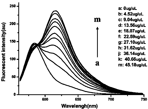
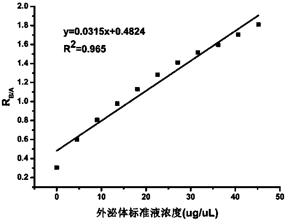
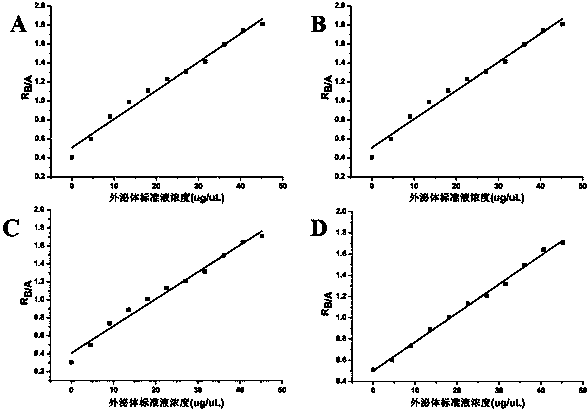
[0063] Take 0, 25, 50, 75, 100, 125, 150, 175, 200, 225, and 250 μL of exosome standard solution, respectively, and make the concentrations of 0, 4.52, 9.04, 13.56, 18.07, 22.59, 27.10, 31.62, 36.14, 40.66, 45.18 μg / μL exosome standard solutions. The PBS solution added to the 0.22μm...


PUM

PropertyMeasurementUnit
diameteraaaaaaaaaa

Abstract

The invention discloses a method for rapidly quantifying exosome by using fluorescence ratio, and belongs to the technical field of fluorescence detection. The method relates to two fluorescent dyes,wherein dye A is a highly water-soluble dye with no affinity to a membrane structure and is used as an internal reference in the experimental process; dye B is a special washing-free membrane dye, andcan show strong luminescence properties after being combined with a lipophilic membrane structure, however, because of the hydrophobicity, the dye B is aggregated without fluorescence in a water environment of not combining with a hydrophilic membrane structure. The invention provides the method for rapidly quantifying the exosome; based on the quantitative ratios of the two fluorescent dyes, thedetermination of the exosomes in a biological sample is successfully realized. The method has the advantages of high efficiency, low cost, convenience in operation, high accuracy of determination results and the like. The method is expected to be widely used in related researches of the exosomes and is applied to cell culture mediums, saliva, serum, urine and emulsion.

Description

technical field [0001] The invention specifically relates to a method for rapid quantification of exosomes using fluorescence ratio, which belongs to the technical field of fluorescence detection. Background technique [0002] Exosomes are small extracellular vesicles formed by cells through the process of "endocytosis-fusion-efflux". Lipid bilayer structure vesicles with a diameter of 30-150nm. As an important intercellular information transfer molecule and genetic material transfer carrier, exosomes contain a variety of proteins and RNA substances, which are widely distributed in human blood, urine, saliva and other body fluids as well as tumor cell culture fluid. Exosomes secreted by tumor cells can promote tumor angiogenesis, and can also affect tumor growth and metastasis by regulating the tumor microenvironment. Exosomes play an important role in the development of tumors. In exosome-related research, it is very important to quickly and accurately quantify the isolat...

Claims


Application Information

Patent Timeline
Patent Type & Authority Applications(China)
IPC IPC(8): G01N21/64
CPCG01N21/6428G01N2021/6439C09B67/0083
Inventor 肖义李宁张新富陈令成
Owner DALIAN UNIV OF TECH
Why Eureka
Try Eureka