Cardiac ultrasound training model

A model and heart technology, applied in the field of medical teaching models, can solve the problems of high price, easy damage to the display screen, and inability to learn, and achieve the effect of good economy and portability

Pending Publication Date: 2018-09-04
PEKING UNION MEDICAL COLLEGE HOSPITAL CHINESE ACAD OF MEDICAL SCI
View PDF6 Cites 2 Cited by
  • Summary
  • Abstract
  • Description
  • Claims
  • Application Information

AI Technical Summary

Problems solved by technology

However, when Sectra Table imports images, it needs to convert CT images into Sectra system anatomical images, which has high requirements for images, because echocardiography training requires images to be images of the circulatory system with enhanced development, including double images of venous phase and arterial phase, so Additional delayed imaging is required for the patient when acquiring images. This inspection sequence is not included in ordinary CT examination items, which not only increases the cost of image acquisition time and patient radiation dose; in the 3D images synthesized with CT-enhanced images, the system cannot The structure and position of the valve are shown well, but the standard echocardiographic view includes several views with the valve as the positioning structure, so it cannot be learned in this image; in addition, the Sectra Table system can only complete the teaching of anatomical cutting in its own machine, but The dissection table is very expensive, and the domestic price is extremely high, so it is difficult to be purchased by ordinary hospitals; in addition, the instrument is large in size and weight, and the display screen is easily damaged. Therefore, long-distance transportation is difficult to achieve, and it cannot meet the needs of a large number of clinical trainings across the country. needs

Method used


Image

  • Cardiac ultrasound training model
  • Cardiac ultrasound training model
  • Cardiac ultrasound training model

Examples


Embodiment 1

[0036] Such as figure 1 As shown, among the present embodiment, the echocardiographic standard section is the parasternal left ventricle long-axis plane 1, and the number of model components is two. Specifically, the model body includes a first model component and a second model component, and the connection plane between the first model component and the second model component is the parasternal left ventricle long axis plane 1. The connecting part plane of the first model component and the second model component is a mirror image structure, and the first model component is the heart bottom part of the heart. The parasternal left ventricle long axis plane 1 includes right ventricle wall 11, right ventricle 32, interventricular septum 12, left ventricle 13, left ventricle inferior posterior wall 14, left ventricle outflow tract, aortic valve 15 and left atrium 16 .

Embodiment 2

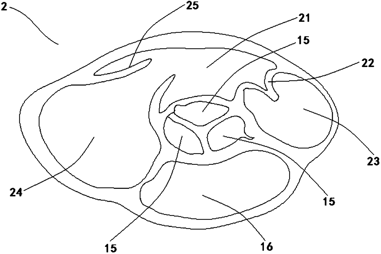
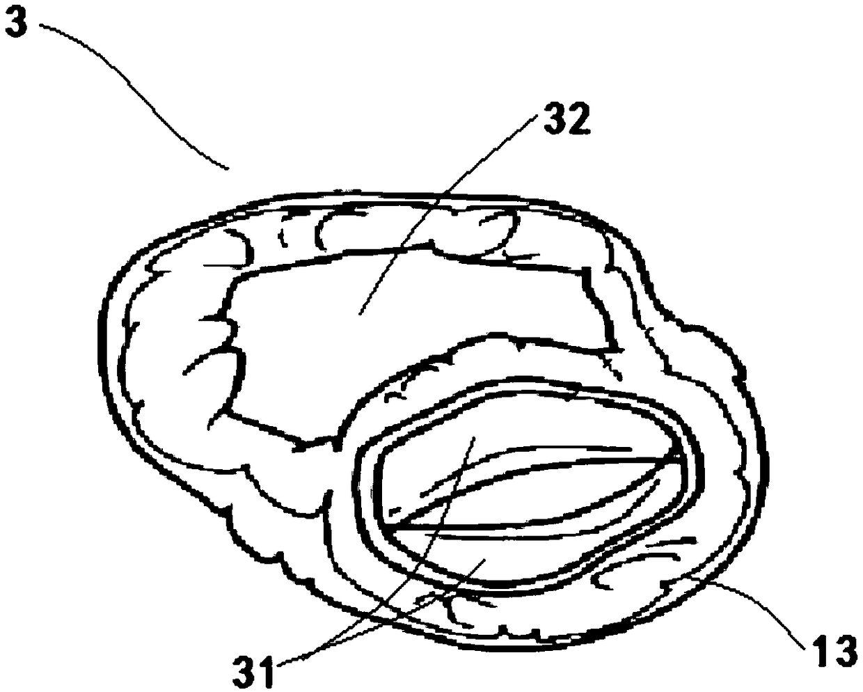
[0038] Such as Figure 2-4 As shown, in this embodiment, the number of model components is four, and the standard echocardiographic views are the parasternal left ventricle short-axis aortic valve plane 2, the parasternal left ventricle short-axis mitral valve plane 3, and the parasternal short-axis mitral valve plane. Axial papillary plane 4. The model main body includes a third model component, a fourth model component, a fifth model component and a sixth model component, wherein the third model component represents the bottom of the heart, the sixth model component represents the apex of the heart, and each model The connections between components are all mirror structures. The connection plane between the third model component and the fourth model component is the parasternal left ventricle short-axis aortic valve plane 2, and the parasternal left ventricle short-axis aortic valve plane 2 contains the aortic valve plane 2 located in the center. The aortic valve 15 and th...

Embodiment 3

[0040] Such as Figure 5-6 As shown, in the present embodiment, the number of model components is three, and the echocardiographic standard slices are the apical five-chamber cardiac plane 5 and the apical four-chamber cardiac plane 6. The main body of the model includes a seventh model component, an eighth model component and a ninth model component. The connection plane between the seventh model component and the eighth model component is the apical five-chamber heart plane 5, and the apical five-chamber heart Plane 5 includes left ventricle 13, mitral valve 31, left ventricular outflow tract, aorta, aortic valve 15, left atrium 16, right ventricle 32, tricuspid valve 25, right atrium 24, interventricular septum 12 and atrial Interval 51; the connection plane between the eighth model component and the ninth model component is the apical four-chamber heart plane 6, which includes the left ventricle 13, the mitral valve valve 31, and the left atrium 16 , right ventricle 32 , ...


PUM


Abstract

The invention relates to a cardiac ultrasound training model comprising a main model body being a model made of a simulated human heart. The main model body consists of a plurality of model assembliesand an acoustic window board assembly. The model assemblies are connected successively from the heart bottom of the main model body to the heat tip; the connection plane between each two adjacent model assemblies is a cardiac ultrasound standard section; and mirror image structures are formed at the connection parts. The acoustic window board assembly has a foldable sector structure; and the multiple model assemblies are connected with the acoustic window board assembly. According to the cardiac ultrasound training model, the main model body is based on the design of the simulated human heartand is formed by successive connection of multiple model assemblies; and with cooperation of adjustment by the acoustic window board assembly, the whole standard section is displayed, so that interpretation transformation from the cardiac anatomy to the ultrasonic image is realized conveniently. Therefore, students are assisted in understand the corresponding relationship of the ultrasound anatomy quickly and thus the teaching goal is achieved quickly. Moreover, the model has advantages of reasonable price, first-rate quality, high portability, high repeatability, and good economic efficiency.

Description

technical field [0001] The invention relates to the technical field of medical teaching models, in particular to a cardiac ultrasound training model. Background technique [0002] Emergency bedside ultrasound training has been widely promoted in China as an emerging project in recent years, and the demand has exploded. With the increase in the amount of training, the quality of training has attracted more and more attention. Bedside ultrasound training Among them, the technical mastery of cardiac color Doppler ultrasound has become a difficult point. Due to certain differences between ultrasound images and solid anatomy, there are certain challenges for students to operate independently in actual operation and in the future for image cognition and understanding. The traditional solid model of the heart is also often used in bedside ultrasound training, and it is often necessary to add pictures for further explanation in order to improve the training effect. The traditional ...

Claims


Application Information

Patent Timeline
IPC IPC(8): G09B23/28
CPCG09B23/286
Inventor 史迪徐军张秋彬朱华栋于学忠
Owner PEKING UNION MEDICAL COLLEGE HOSPITAL CHINESE ACAD OF MEDICAL SCI
Why Eureka
Try Eureka