A simulation system for simulating fracture healing process
A fracture healing and simulation system technology, applied in the field of biomedical engineering, can solve the problems of not describing the angiogenesis process of fracture healing from different levels, not accurately establishing cell concentration, and not having fracture healing, so as to avoid humanitarian controversy and reduce Effect of delaying union and reducing fracture nonunion
- Summary
- Abstract
- Description
- Claims
- Application Information
AI Technical Summary
Problems solved by technology
Method used
Image
Examples
specific Embodiment approach 1
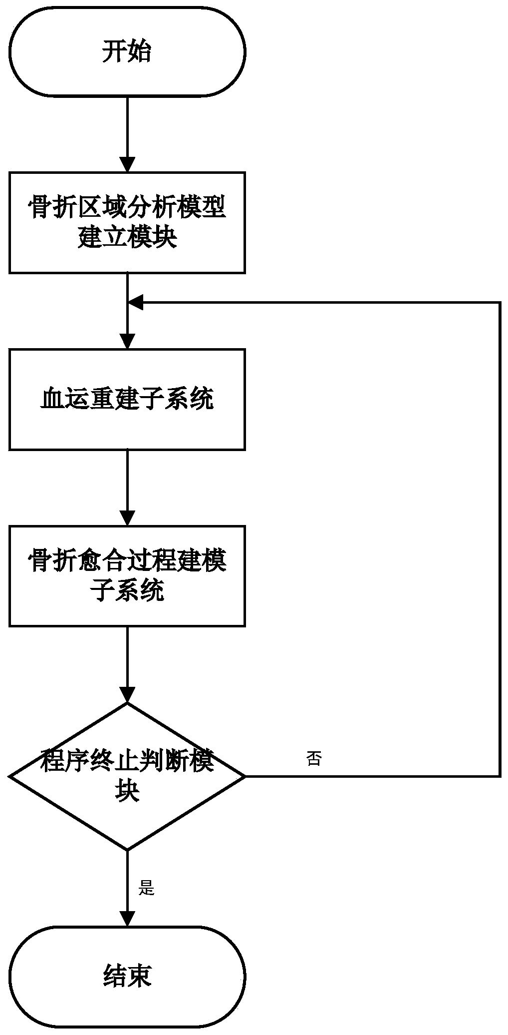
[0036] Specific implementation mode one: as figure 1 As shown, a simulation system for simulating the fracture healing process described in this embodiment includes:
[0037] Fracture area analysis model establishment module 1, revascularization subsystem 2, fracture healing process modeling subsystem 3 and procedure termination judgment module 4;
[0038] Fracture area analysis model establishment module 1 is used to establish fracture area geometric model and finite element analysis model;
[0039] The revascularization subsystem 2 includes:
[0040] The modeling module of intracellular molecular physiological activities is used to model the revascularization process during fracture healing from the relevant molecular physiological activities at the intracellular molecular level;
[0041] The related molecules include angiogenesis cell growth factor receptor, Notch1 protein, Dll4 protein, activated angiogenesis cell growth factor receptor, activated Notch1 protein, effecti...
specific Embodiment approach 2
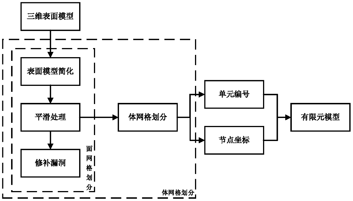
[0052] Specific implementation mode two: as Figure 1-4 As shown, in this embodiment, the specific process of the fracture area analysis model building module 1 to realize its function is:
[0053] 1) The establishment of a three-dimensional surface geometric model of the fracture area;
[0054] Using the segmentation-based 3D medical image surface reconstruction algorithm to reconstruct the surface of the image, and obtain the 3D surface geometric model through the process of threshold screening, interactive segmentation and 3D reconstruction;
[0055] The image is obtained by imaging equipment CT, and the data storage format is DICOM;
[0056] 2) Establishment of the finite element model of the fracture area;
[0057] Mesh the three-dimensional surface geometric model of the fracture area, discretize the continuous geometric model, and obtain the finite element model of the fracture area;
[0058] The meshing includes two steps of surface meshing and volume meshing: the sur...
specific Embodiment approach 3
[0063] Specific implementation mode three: as Figure 1-4 As shown, in this embodiment, the specific process for the revascularization subsystem 2 to realize its functions is as follows:
[0064] 1)Molecular physiological activity modeling module inside the cell
[0065] The process of angiogenic growth factor receptor activation is described as follows:
[0066]
[0067] In the formula, V t 'is the number of activated angiogenic growth factor receptors at time t, V sink is the number of angiogenic cell growth factor decoy receptors, t is time, δt is the cycle time of the subroutine, V t-δt is the number of angiogenic cell growth factor receptors at time t-δt, V max is the maximum number of angiogenic cell growth factor receptors, g vessel is the concentration of angiogenic cell growth factor, M tot is the total amount of angiogenesis cell membrane;
[0068] The modeling process of Dll4 protein quantity is as follows:
[0069] D. t =D t-δt +V″ t-δt δ-N' t-δt,nei...
PUM
 Abstract
Description
Claims
Application Information
 - R&D
- Intellectual Property
- Life Sciences
- Materials
- Tech Scout
- Unparalleled Data Quality
- Higher Quality Content
- 60% Fewer Hallucinations
Browse by: Latest US Patents, China's latest patents, Technical Efficacy Thesaurus, Application Domain, Technology Topic, Popular Technical Reports.
© 2025 PatSnap. All rights reserved.Legal|Privacy policy|Modern Slavery Act Transparency Statement|Sitemap|About US| Contact US: help@patsnap.com


