A Method for Retinal Layering in Fundus OCT Images
An image, retinal technology, applied in the field of medical image processing, to achieve good repeatability and adaptability, verification accuracy and reliability, and strong comparability.
- Summary
- Abstract
- Description
- Claims
- Application Information
AI Technical Summary
Problems solved by technology
Method used
Image
Examples
Embodiment Construction
[0054] The present invention will be described in further detail below in conjunction with the accompanying drawings and embodiments. It should be understood that the specific embodiments described here are only used to explain the present invention, not to limit the present invention.
[0055] Embodiments of the present invention are as follows:
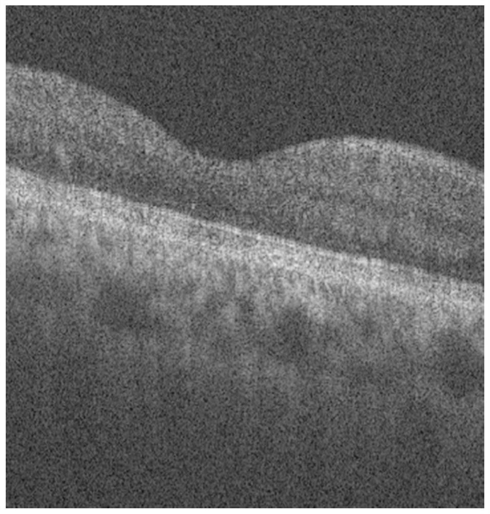
[0056] 1) Collect the OCT image of the fundus retina as the original image, such as figure 2 shown.
[0057] 2) Use the weight coefficient matrix template [1 / 9, 1 / 9, 1 / 9; 1 / 6, 1 / 6, 1 / 6; 1 / 9, 1 / 9, 1 / 9;] to traverse the entire original image for Template filtering;
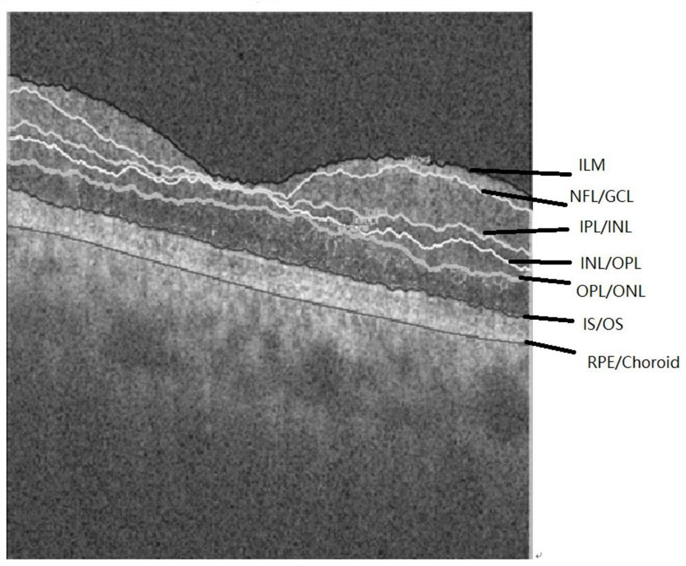
[0058] 3) Process in the following manner to obtain each boundary line, and perform retinal delamination in the fundus OCT image.
[0059] 3.1) For each column of the OCT image, record the pixel point where the maximum gray value is located, and connect the pixel points where the maximum gray value of each column is located as the dividing line between the retinal pi...
PUM
 Abstract
Description
Claims
Application Information
 - R&D
- Intellectual Property
- Life Sciences
- Materials
- Tech Scout
- Unparalleled Data Quality
- Higher Quality Content
- 60% Fewer Hallucinations
Browse by: Latest US Patents, China's latest patents, Technical Efficacy Thesaurus, Application Domain, Technology Topic, Popular Technical Reports.
© 2025 PatSnap. All rights reserved.Legal|Privacy policy|Modern Slavery Act Transparency Statement|Sitemap|About US| Contact US: help@patsnap.com


