Eureka AIR delivers breakthrough ideas for toughest innovation challenges, trusted by R&D personnel around the world.

Method for promoting human cholangiocytes to differentiate and mature through MAPK/PKC signaling pathway activator

A signal pathway and activator technology, applied in the field of MAPK/PKC signal pathway activator to promote the differentiation and maturation of human cholangiocytes, can solve the problem of not fully reflecting the physiological and pathological characteristics of bile ducts in vivo, limiting the scope of application, reliability, and expression level low level problem

Active Publication Date: 2018-11-23
PEKING UNIV +1
View PDF5 Cites 6 Cited by
  • Summary
  • Abstract
  • Description
  • Claims
  • Application Information

AI Technical Summary

Problems solved by technology

However, these intrahepatic cholangioid cell induction protocols and the induced intrahepatic cholangioid cells have the following disadvantages, which also limit the scope and reliability of their application
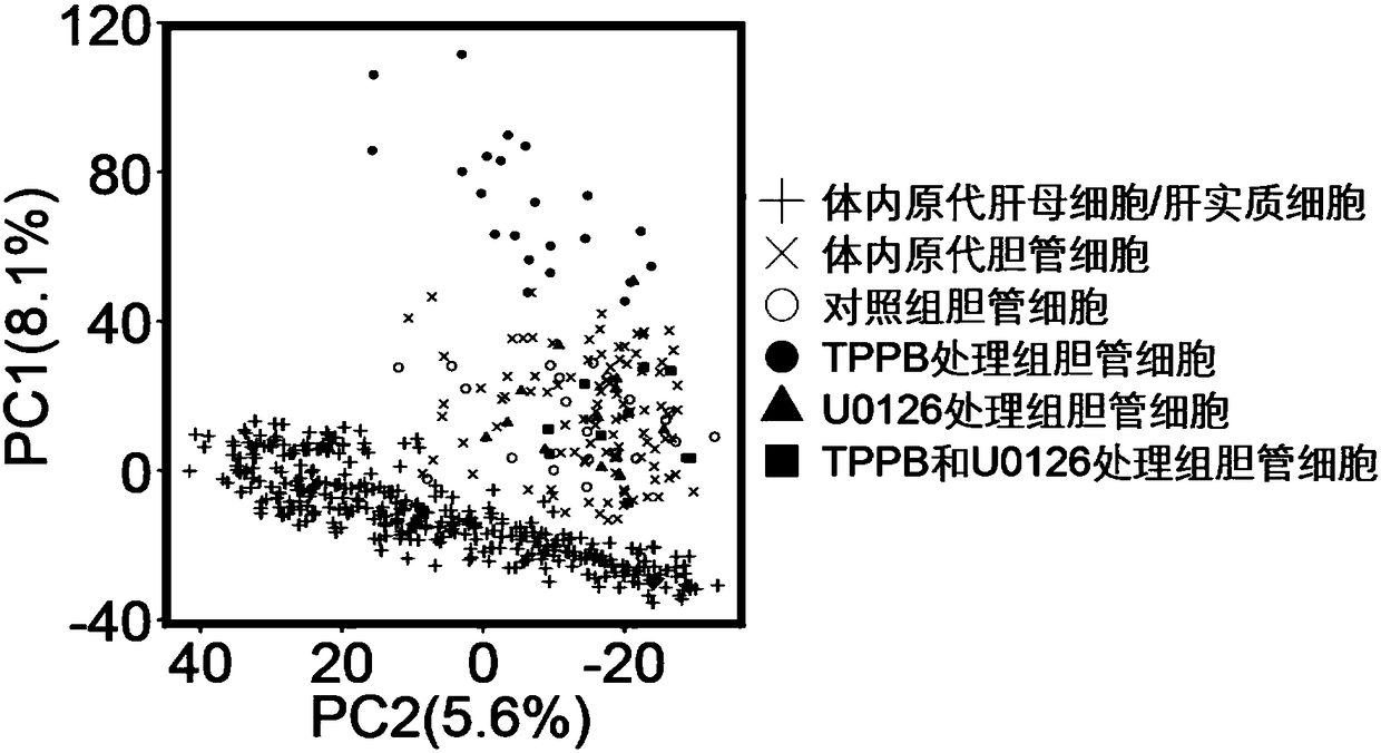
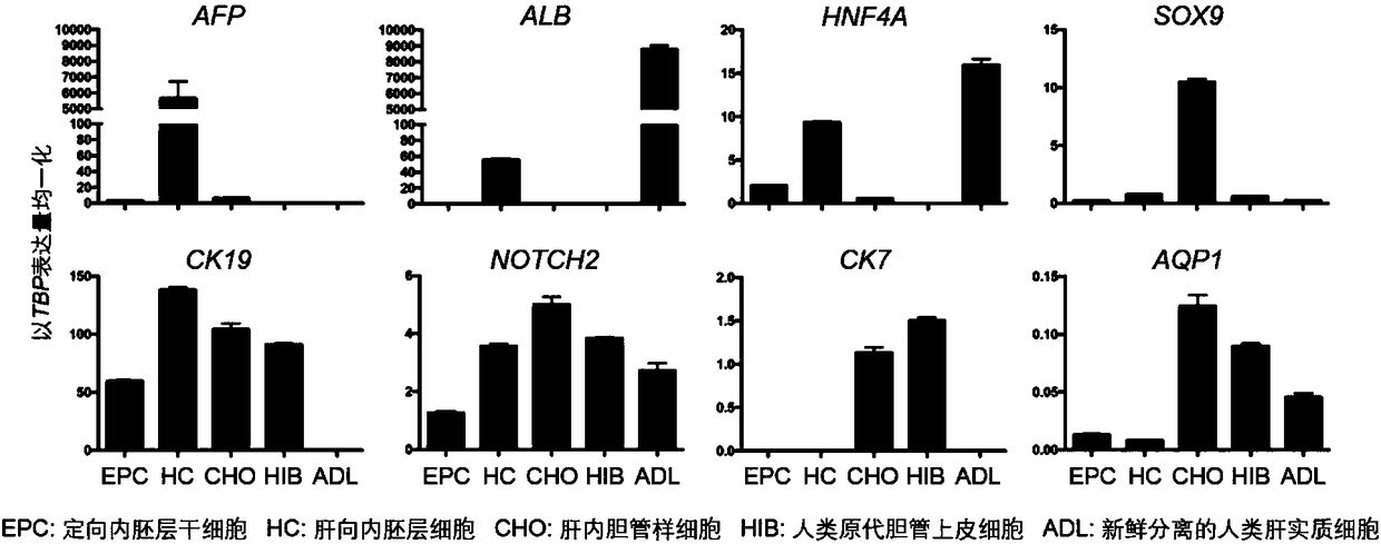
[0005] (1) The differentiation efficiency and purity of cholangioid cells are low, the induction time is long (>26 days), and the cost is high; (2) The gene expression profiles of cholangioid cells and mature intrahepatic cholangiocytes from adult liver Significant differences exist, with high expression of marker genes of immature intrahepatic cholangiocytes, and low or no expression of genes related to mature intrahepatic cholangiocytes; (3) Bile duct-like cells and mature intrahepatic cholangiocytes from adult liver Compared with cholangiocytes, its function is not perfect and immature, including low enzyme activity (alkaline phosphatase (ALP) and γ-glutamyl transpeptidase (γ-GGT), etc.), low CFTR-mediated chloride Compound transport activity and limited hormone (secretin and somatostainin) response activity, etc., indicate that cholangioid cells are immature; (4) Due to the different types of small molecule compounds used in the existing in vitro induction schemes of intrahepatic cholangioid cells, the The order of addition and treatment time are also different, and some programs even need to add trophoblast cells, indicating that these programs do not completely simulate the normal development of intrahepatic bile ducts, and the induced differentiation of bile duct-like cells does not fully reflect the Physiological and pathological characteristics of bile ducts in vivo; (5) bile duct-like cells lack functional verification in vivo (model animals and humans)

Method used


Image

  • Method for promoting human cholangiocytes to differentiate and mature through MAPK/PKC signaling pathway activator
  • Method for promoting human cholangiocytes to differentiate and mature through MAPK/PKC signaling pathway activator
  • Method for promoting human cholangiocytes to differentiate and mature through MAPK/PKC signaling pathway activator

Examples


Embodiment 1

[0031] Example 1: The effect of TPPB on the differentiation and maturation of mouse intrahepatic cholangiocytes

[0032] 1) Differentiation and development of mouse intrahepatic cholangiocytes

[0033] C57BL / 6-strain female mice and C3H-strain male mice from 8 to 12 weeks old came from Victoria Lihua Company (Beijing), and offspring mice (B6C3F1) obtained from mating were used for subsequent experiments. B6C3F1 female mice of about six weeks are mated with B6C3F1 male mice of eight to twelve weeks, and the vaginal suppository is found in the next morning, which is recorded as embryonic development (E) 0.5 days. All mice were housed in a sterile environment at 23 ± 2°C for 12 hours with day and night cycles, and fed with autoclaved food and water. All mouse experiments were carried out in strict accordance with the management regulations of the Experimental Animal Center of Peking University.

[0034] Take liver buds or fetal livers at 10.5, 11.5, 12.5, 13.5, 14.5, 15.5, and ...

Embodiment 2

[0039] Example 2: Preparation and maintenance of human committed endoderm stem cells

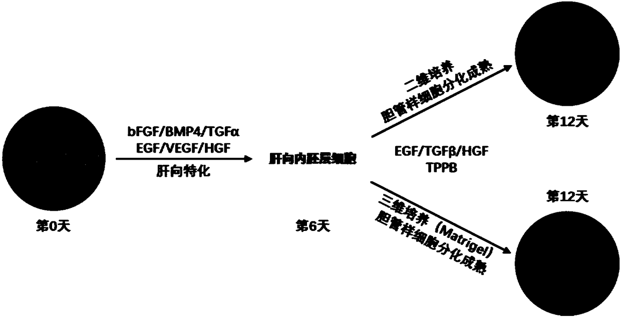
[0040] Committed endoderm stem cells (EP cells) were prepared from human induced pluripotent stem cells (iPSCs) using the method disclosed in WO 2012 / 178215 Al by Gadue et al. Human induced pluripotent stem cells (iPSCs) (purchased from Shanghai Zhongqiao Xinzhou Biotechnology Co., Ltd.) were treated with glutamine (2 mM), MTG (4.5×10 -4 M), activin A (100ng / ml) and CHIR99021 (2M) in the RPMI medium induced for 1 day, followed by glutamine (2mM), ascorbic acid (50g / ml), MTG (4.5 × 10 -4 M), bFGF (5ng / ml) and activin A (100ng / ml) in the RPMI medium for directional induction for 2 days, and finally in the RPMI medium containing glutamine (2mM), ascorbic acid (50g / ml), MTG (4.5×10 -4 M), bFGF (5 ng / ml) and activin A (100 ng / ml) were induced in SFD medium for 2 days. The CXCR4+ / CD117+ cells were sorted out by flow cytometry to obtain human committed endoderm stem cells.

[0041] The resulting...

Embodiment 3

[0042] Example 3: Directed differentiation of endoderm stem cells from liver to endoderm

[0043] Committed endoderm stem cells were differentiated into the liver by suspension culture, and the cell density was 1 million / ml differentiation medium (containing BMP4 (50ng / ml), bFGF (10ng / ml), VEGF (10ng / ml), EGF (10ng / ml) ml), TGFα (10ng / ml), HGF (25ng / ml), Dex (40ng / ml), glutamine (2mM), ascorbic acid (50g / ml) and MTG (4.5×10 -4 M) SFD medium), add 100μl matrix collagen solution and pre-cooled cell suspension to each 3ml medium, mix well, place in a low-adsorption culture dish for directional differentiation, the differentiation time is 6 days, and replace every 2 days medium once.


PUM


Abstract

The invention provides a method for inducing directional endoderm stem cells to form intrahepatic bile duct-like cells. The method comprises the steps of directionally inducing directional endoderm stem cells to form liver directed endodermal cells, and differentiating the liver directed endodermal cells into human bile duct-like cells under directional action of a small molecular compound and theMAPK / PKC signaling pathway activator. The invention further provides human bile duct-like cells acquired through the method.

Description

technical field [0001] The invention relates to a method for inducing differentiation of human pluripotent stem cells to form intrahepatic bile duct-like cells. In particular, the present invention relates to a method for directionally inducing human induced pluripotent stem cells into committed endoderm stem cells, and further inducing to form intrahepatic bile duct-like cells. Background technique [0002] Due to the needs of pathological research, drug screening and disease treatment of intrahepatic bile duct diseases, it is extremely important and urgent to efficiently induce human pluripotent stem cells to obtain mature and functional intrahepatic bile duct-like cells. [0003] At present, several research groups have successively published protocols for inducing the differentiation of human pluripotent stem cells to form intrahepatic bile duct-like cells. Sampaziotis F et al. started from human induced pluripotent stem cells (hiPSCs) and induced them to form committed...

Claims


Application Information

Patent Timeline
IPC IPC(8): C12N5/071C12N5/10
CPCC12N5/067C12N2501/11C12N2501/115C12N2501/12C12N2501/148C12N2501/155C12N2501/727C12N2501/734C12N2506/45
Inventor 徐成冉程新杨李冯思思吴佳颖邱伟林
Owner PEKING UNIV
Why Eureka
Eureka Blog
Learn More