Method for quickly preparing hard tissue slice and application of the method

A hard tissue and slicing technology, applied in the field of biomedicine, can solve the problems of complex preparation process, long time-consuming and high cost, and achieve the effects of simple and easy operation, short time-consuming and low cost.

Active Publication Date: 2018-11-30
SUZHOU UNIV
View PDF8 Cites 6 Cited by
  • Summary
  • Abstract
  • Description
  • Claims
  • Application Information

AI Technical Summary

Problems solved by technology

[0004] In order to solve the above-mentioned technical problems, the object of the present invention is to provide a method for rapidly preparing hard tissue slices and its application, which overcomes the complicated preparation process, high cost and long time-consuming (need 2-3 weeks) of traditional hard tissue slice methods. Disadvantages, provide an easy-to-learn, cheap, and fast (only 3 days) preparation method for hard tissue slices

Method used


Image

  • Method for quickly preparing hard tissue slice and application of the method
  • Method for quickly preparing hard tissue slice and application of the method
  • Method for quickly preparing hard tissue slice and application of the method

Examples


Embodiment 1

[0051] Example 1 Preparation of tibial hard tissue slices of 3-month-old mice

[0052] 1. Mouse tibia tissue acquisition, fixation, embedding and quick freezing in liquid nitrogen

[0053](1) Execute the mice, take the tibia, remove excess soft tissue, place in pre-cooled 4% paraformaldehyde (PFA), store and fix at 4°C overnight;

[0054] (2) Take out the fixed sample, wash it three times with PBS, each time for 5 minutes, then soak it in 15% (w / v) sucrose solution for 1 hour, then transfer to 30% (w / v) sucrose overnight at 4°C in the solution;

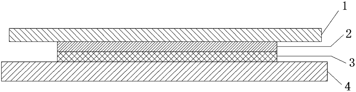
[0055] (3) Place the sucrose-soaked sample in the embedding box containing OCT embedding agent, and adjust the sample to a suitable orientation, and mark the position where sectioning is required;

[0056] (4) Take a cell culture dish with a diameter of 10 cm and place it in a foam box containing liquid nitrogen, so that the culture dish floats on the liquid nitrogen and pre-cool for 1 minute, and pay attention to avoid liquid nitro...

Embodiment 2

[0080] Example 2 Preparation of 3-month-old rat femoral hard tissue slices

[0081] The mouse in Embodiment 1 was replaced with a rat, and its femur was taken, and other steps were carried out according to Embodiment 1 to prepare rat femoral hard tissue slices.

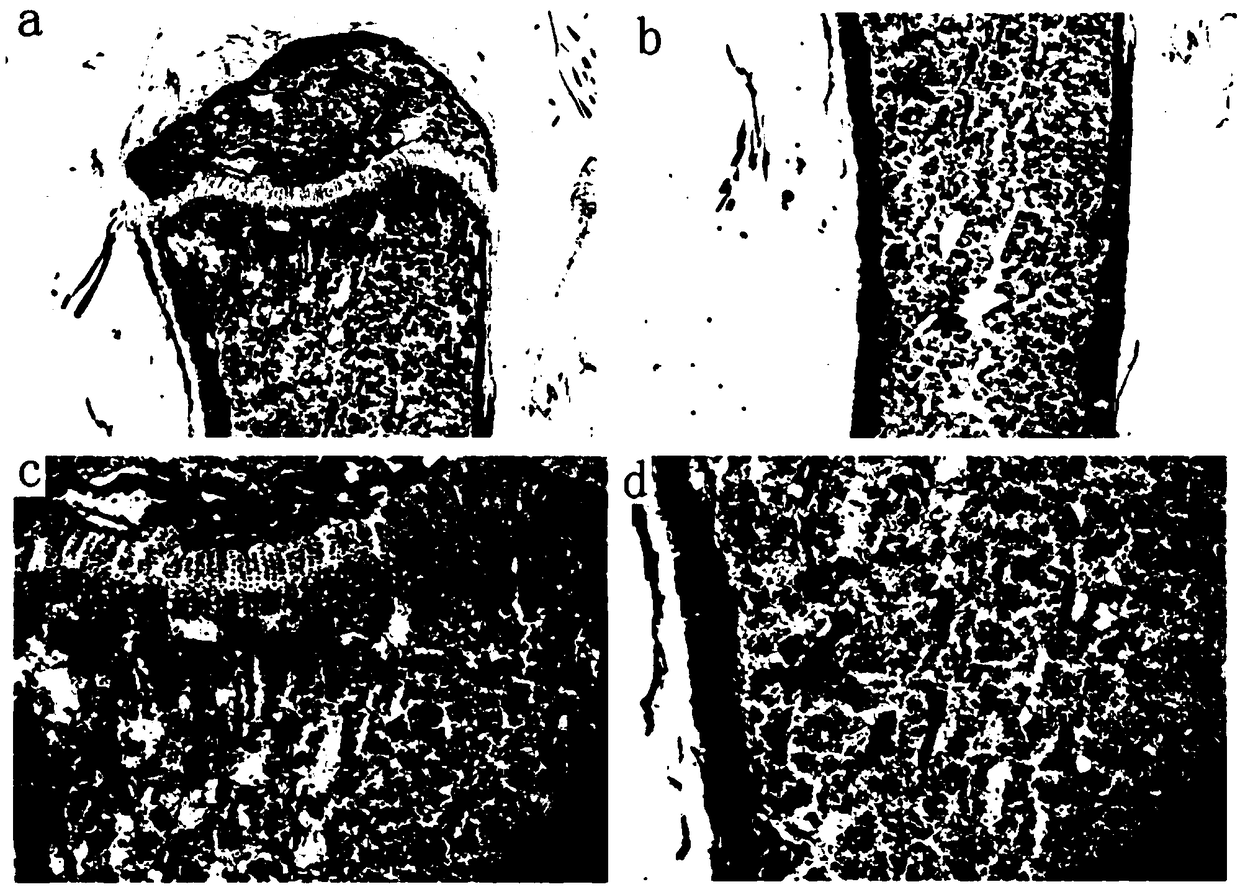
[0082] According to the method of embodiment 1, the calcein fluorescence color double labeling is carried out to rat femur hard tissue section, the result is as follows Figure 4 as shown, Figure 4 a-d represent fluorescence micrographs at different positions of the rat femur, respectively. In the figure, DAPI-labeled cell nuclei appear blue, and calcein fluorescently-labeled newborn bone appears green. The results show that the rat femur slices obtained by this method have complete bone Comparing with the bone marrow structure, the calcein fluorescently labeled new bone is clearly visible, which can fully satisfy the downstream experimental analysis.


PUM

PropertyMeasurementUnit
thicknessaaaaaaaaaa

Abstract

The invention relates to a method of quickly preparing a hard tissue slice, which includes the steps of: 1) immobilizing the hard tissue in paraformaldehyde and soaking the hard tissue in a sucrose water solution; 2) embedding the hard tissue with an embedding agent and rapid-freezing the hard tissue to cure the embedding agent; 3) tightly adhering one side of the sample to an adhesion film strip,and slicing the hard tissue at -25 to -30 DEG C in the direction being parallel with the surface of the adhesion film strip, slicing thickness being 6-8 [mu]m; 4) placing the film strip, with the hard tissue slice, on an anti-stripping glass slide, tightly adhering the hard tissue slice on the film strip with the anti-stripping glass slide through ultraviolet curable glue, then initiating the polymerization of the ultraviolet curable glue under ultraviolet light and removing the adhesion film strip on the surface of the hard tissue slice, thus obtaining the hard tissue slice. The invention also claims to protect application of the hard tissue slice, prepared through the method, in tissue staining. The method is easy to master and is low-cost and quick (only need three days).

Description

technical field [0001] The invention relates to the field of biomedicine, in particular to a method for rapidly preparing hard tissue slices and an application thereof. Background technique [0002] Hard tissue (bones, teeth, etc.) diseases, especially orthopedic diseases such as osteoporosis, are common and serious diseases. Tissue section technology is an important means to study the pathological process. The traditional method of slicing hard tissues such as bone is to fix, decalcify, embed and slice the hard tissues, and then stain them for observation. However, during the decalcification process, some important proteins are often denatured and some important signals are lost, so that sensitive indicators are difficult to detect, which seriously affects the experimental results. In addition, some important indicators (such as calcification status, dynamic parameters of bone formation, etc.) must be observed through hard tissue sections. At present, the most commonly u...

Claims


Application Information

Patent Timeline
Patent Type & Authority Applications(China)
IPC IPC(8): G01N1/28
CPCG01N1/286G01N2001/2873
Inventor 陈建权郑懿欣刘存昌
Owner SUZHOU UNIV
Why Eureka
Try Eureka