Application of GMFB antibody in preparation of drugs for treating diabetic retinopathy

A technology for treating diabetic retina and drug, which is applied in the application field of GMFB antibody as a drug for preparing diabetic retinopathy, can solve the problems of decreased visual sensitivity and color vision sensitivity, affecting the progress of DR, abnormality and the like, and achieves the improvement of visual function. Effect

Pending Publication Date: 2018-12-07
TONGJI UNIV
View PDF1 Cites 7 Cited by
  • Summary
  • Abstract
  • Description
  • Claims
  • Application Information

AI Technical Summary

Problems solved by technology

Schellin SA et al. used light microscopy and electron microscopy to detect that Muller cell nuclei had changed in the early stage of DM, while retinal vascular endothelial cells and pericytes had no obvious pathological changes. The ultrastructure and physiological functions of retinal Muller cells in the early stage of DM had changed. It not only affects the abnormal function of nerve cells in patients with early DM (shown as decreased visual sensitivity and color sensitivity, and abnormal b-wave of retinal oscillatory potential), but also affects the progress of the entire DR

Method used


Image

  • Application of GMFB antibody in preparation of drugs for treating diabetic retinopathy
  • Application of GMFB antibody in preparation of drugs for treating diabetic retinopathy
  • Application of GMFB antibody in preparation of drugs for treating diabetic retinopathy

Examples


Embodiment 1

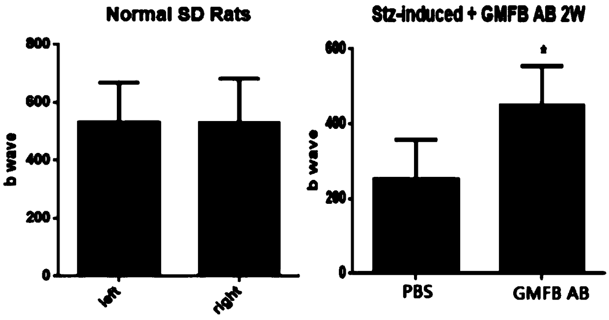
[0036] Example 1: Intravitreal injection of GMFB antibody improves visual function in DR rats.

[0037] (1) Preparation of diabetic rats: male SD rats weighing 160-180 g were used, and the rats were starved for 24 hours before the experiment. A single intraperitoneal injection of STZ (60mg / kg body weight) to induce DM, and an equal volume of citric acid solution was injected intraperitoneally in the normal control group; 24 hours later, blood was taken from the tail to measure blood glucose, and rats with blood glucose values ​​lower than 250mg / dL were given supplementary injections STZ. Blood sugar was measured for 3 consecutive days. Rats with blood glucose exceeding 250 mg / dL for 3 consecutive days were determined as DM rats (rats with blood glucose lower than 250 mg / dL would be excluded).

[0038] (2) GMFB antibody injection: On the day of onset of STZ-TIDM rats, 10 ul of GMFB antibody (concentration: 10 ug / 150 ul.) was injected into the vitreous cavity of the right eye, a...

Embodiment 2

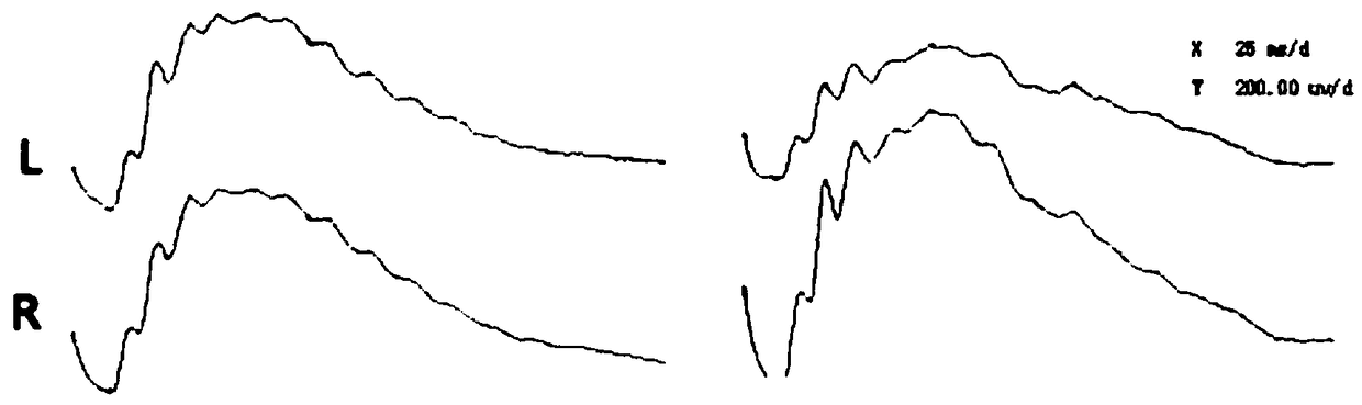
[0039] Example 2: ERG detection.

[0040] APS Automatic Visual Electrophysiology Tester (APS-2000) was purchased from Chongqing Kanghua Technology Co., Ltd.

[0041] (1) The day before the visual electrophysiological function test, the DM rats were transferred to a dark room for dark adaptation.

[0042] (2) Preparation of rats: intraperitoneal injection of 2% sodium pentobarbital (1mL / 500g body weight) to the rats for anesthesia, 1× Sumianxin (0.1ml / 200g) to make the eyeballs protrude, and then give a drop of 0.5% tonbarbital Mydriasis was performed with picamide (Wuxi Shanhe Group, Jiangsu, China), a drop of 0.4% oxybucaine hydrochloride was used for topical anesthesia (Eisai Co Ltd, Tokyo, Japan), and a little conductive paste was applied to each eye.

[0043] (3) Insert electrodes: connect the ground wire to the tail of the rat, connect the negative electrode between the two ears of the rat, and connect the positive electrode to the cornea of ​​both eyes, and be careful n...

Embodiment 3

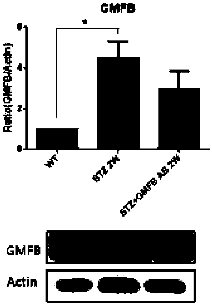
[0046] Example 3: Detection of protein expression level in retina by Western Blot.

[0047] (1) Wash the retina twice with PBS buffer, add 200ul RIPA lysate, and use a homogenizer to lyse the retinal tissue into single cells.

[0048] (2) Place it on ice for 30 hours to fully lyse, and vortex for a few seconds every ten minutes.

[0049] (3) Centrifuge at 12000 rpm for 15 minutes at 4°C.

[0050] (4) Load the sample with a protein amount of 30ug / well, and then perform SDS-PAGE electrophoresis, electrophoresis conditions: 90V, 30min; 120V, until the end.

[0051] (5) Transmembrane transfer of protein to PVDF membrane. The condition is 300mA, 2h.

[0052] (6) Use rabbit-derived GMFB, GFAP antibody and anti-rabbit HRP-labeled secondary antibody to detect the expression of the corresponding protein, and actin is used as an internal reference.

[0053] At 2 weeks after intravitreal injection of GMFB antibody, the retina was taken to detect the expression of GMFB and GFAP by Wes...


PUM


Abstract

The invention relates to an application of a GMFB antibody, in particular, an application of the GMFB antibody in the preparation of drugs for treating diabetic retinopathy. The GMFB antibody is injected into the right eye of an STZ induced DM rat by a vitreous cavity injection technology, then PBS with an equal amount is injected into the left eye, after two weeks, the difference of visual functions of two eyes of the rat is detected by an ERG method, and the result shows that the visual function of the eyeball that is injected with the GMFB antibody is obviously improved, compared with thatof the eyeball that is not injected with the GMFB antibody. Then people find that the GFAP expression of the retina of the eyeball with the GMFB antibody is prominently reduced, TUNEL dyeing for detecting cell apoptosis is also weakened, and the result shows that the GMFB antibody injected into the vitreous cavity can be used as a target of DR early intervention to protect the visual function.

Description

technical field [0001] The present invention relates to the application of the antibody GMFB, in particular to the application of the GMFB antibody as a drug for the preparation of diabetic retinopathy. Background technique [0002] With the rapid development of my country's economy and the acceleration of the aging process, the prevalence of diabetes (diabetes milletus, DM) is showing a rapid upward trend, becoming another important chronic disease that seriously endangers people's health after cardiovascular and cerebrovascular diseases and tumors. non-communicable diseases. The World Health Organization predicts that in 2025, the number of diabetic patients in China will reach 300 million. Diabetic Retinopathy (DR), referred to as Diabetic Retinopathy, is the most common complication of diabetes. DR occurs in 1 / 3 of DM patients, and fatal vision-threatening diabetic macular edema (DME) occurs in 1 / 10. ) or proliferative diabetic retinopathy (PDR), seriously affecting the...

Claims


Application Information

Patent Timeline
Patent Type & Authority Applications(China)
IPC IPC(8): A61K39/395A61P27/02A61P3/10
CPCA61K2039/505A61P3/10A61P27/02C07K16/18
Inventor 吕立夏徐国彤朱彤张介平王娟
Owner TONGJI UNIV
Why Eureka
Try Eureka