Method for acquiring macrophages with phagocytic functions by means of pluripotent stem cell differentiation
A technology of grouping cells and cytokines, applied in the direction of non-embryonic pluripotent stem cells, artificially induced pluripotent cells, biochemical equipment and methods, etc., can solve the problem of immature iPS induction differentiation method, few sources of macrophages, and poor quality Guarantee and other issues to achieve the effect of low cost, good growth, and reduced pollution risk
- Summary
- Abstract
- Description
- Claims
- Application Information
AI Technical Summary
Problems solved by technology
Method used
Image
Examples
Embodiment 1
[0133] Example 1 Induction of hiPSCs to form embryoid bodies (EBs)
[0134] hiPSCs are obtained from human peripheral blood mononuclear cells (PBMC) through the existing method of inducing reprogramming: a certain amount of PBMCs are electroporated into episomes containing reprogramming genes, and after clones appear, they are picked on matrigel culture plates to continue culturing , to obtain reprogrammed iPS.
[0135] Prewarm (15-25°C) sufficient amounts of mTeSR1, DMEM / F12 and Versene for cell passage. The Rock kinase inhibitor Y-27632 was added to the medium so that the final concentration was 3 μM.
[0136] a) Wash the original well with 1ml DPBS;
[0137] b) Absorb DPBS, add 1ml Versene+Y27632;
[0138] c) Incubate at 37°C for 4 minutes;
[0139] d) Use a pipette with a 1000ul tip to pipette 1-2 times and remove the cells (usually the cells are still in larger clumps to form better EBs);
[0140] e) Immediately transfer the cells to a centrifuge tube containing DMEM...
Embodiment 2
[0142] Example 2 Induction of embryoid bodies (EBs) to differentiate into macrophages
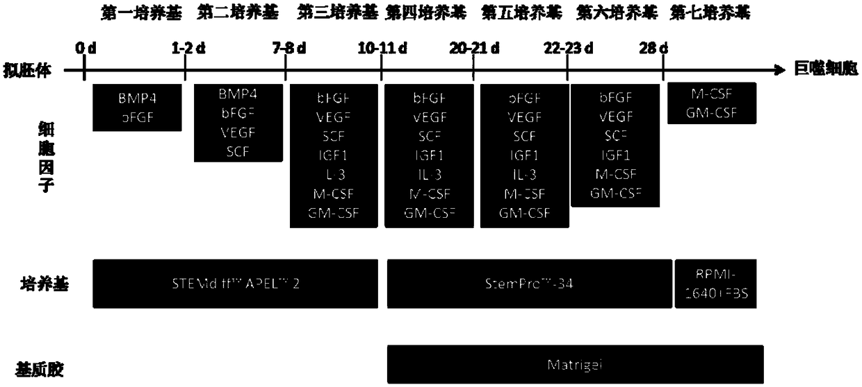
[0143] Schematic flow chart of embryoid body (EB) induced differentiation into macrophages as shown in figure 1 shown.
[0144] Step a) remove the mTeSR1 medium of the embryoid body in f) of embodiment 1, and use the first medium (STEMdiff TM APEL TM 2. 10ng / ml BMP4, 5ng / ml bFGF) were incubated for 24 hours, and the embryoid bodies were differentiated into mesoderm cells;
[0145] Step b) remove the first culture medium in step a), start to use the second culture medium (STEMdiff TM APEL TM 2, 10ng / ml BMP4, 5ng / ml bFGF, 50ng / ml VEGF, 100ng / ml SCF) incubate and culture mesoderm cells to obtain hematopoietic cells, during which a new second medium is replaced every other day;
[0146] Step c) remove the second culture medium in step b), start to use the third culture medium (STEMdiff TM APEL TM 2, 10ng / ml bFGF, 50ng / ml VEGF, 50ng / ml SCF, 10ng / ml IGF1, 25ng / ml IL-3, 50ng / ml M-CSF, 50ng / ...
Embodiment 3
[0152] Example 3 Detection of Cell Differentiation Morphology
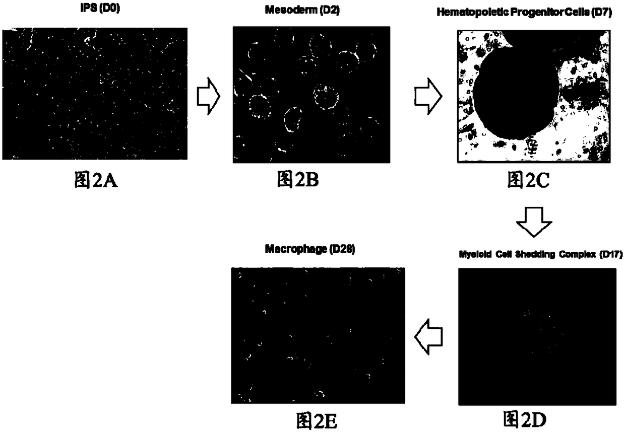
[0153] Observe the cell morphology under a microscope and take pictures, the results are as follows Figure 2A-Figure 2E shown.
[0154] Figure 2A For iPS cells on day 0, Figure 2B For day 2 mesoderm cells, Figure 2C For day 7 hematopoietic cells, Figure 2D For myeloid cells on day 17, Figure 2E Mature macrophages on day 28.
[0155] It can be seen from the figure that the iPS cells were well differentiated and a large number of mature macrophages were obtained.
PUM
 Abstract
Description
Claims
Application Information
 - R&D
- Intellectual Property
- Life Sciences
- Materials
- Tech Scout
- Unparalleled Data Quality
- Higher Quality Content
- 60% Fewer Hallucinations
Browse by: Latest US Patents, China's latest patents, Technical Efficacy Thesaurus, Application Domain, Technology Topic, Popular Technical Reports.
© 2025 PatSnap. All rights reserved.Legal|Privacy policy|Modern Slavery Act Transparency Statement|Sitemap|About US| Contact US: help@patsnap.com


