Aptamer group for detecting exosomes, lateral flow aptamer biosensor and preparation method thereof

A biosensor and aptamer technology, applied in biochemical equipment and methods, biological testing, instruments, etc., can solve the problems of complex steps, expensive synthesis, denaturation, etc., and achieve high specificity and easy operation

Active Publication Date: 2019-02-12
南通拓普赛斯生物科技有限公司
View PDF2 Cites 10 Cited by
  • Summary
  • Abstract
  • Description
  • Claims
  • Application Information

AI Technical Summary

Problems solved by technology

Although the use of antigen-antibody immunoassay can achieve high detection sensitivity and does not require large-scale instruments, the screening of antibodies that specifically bind to exosome surface marker proteins is often complicated, and antibody proteins are easily affected by environmental factors such as pH and temperature. Denaturing and expensive to synthesize

Method used


Image

  • Aptamer group for detecting exosomes, lateral flow aptamer biosensor and preparation method thereof
  • Aptamer group for detecting exosomes, lateral flow aptamer biosensor and preparation method thereof
  • Aptamer group for detecting exosomes, lateral flow aptamer biosensor and preparation method thereof

Examples


preparation example Construction

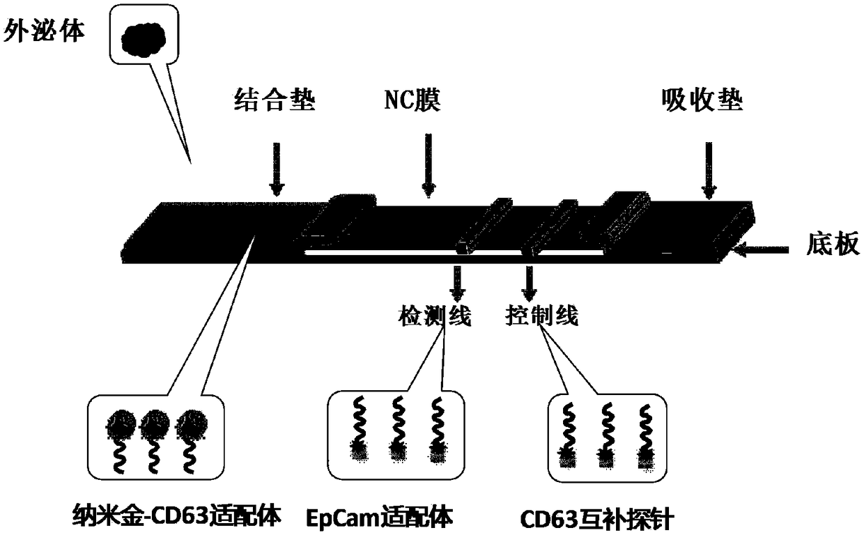
[0042] The invention provides a method for preparing a lateral flow aptamer biosensor, comprising the following steps:

[0043] (1) Add dATP solution, SDS solution and NaCl solution to the nano-gold solution under shaking conditions to react, and the obtained reaction solution is mixed with the CD63 aptamer, reacted at 60°C for 3 hours, separated from the solid and liquid, and the obtained precipitate Dissolving in the nanoparticle storage solution to obtain a gold nanometer-labeled CD63 aptamer conjugate solution and spraying it on the binding pad to obtain a binding pad sprayed with the gold nanometer-labeled CD63 aptamer conjugate;

[0044] (2) After incubating the biotin-labeled EpCam aptamer and streptavidin for 1 hour, place it in an ultrafiltration centrifuge tube for ultrafiltration centrifugation, wash the ultrafiltration centrifuge tube twice, and collect the supernatant as follows: Streptavidin-biotin-labeled EpCam aptamer solution;

[0045] (3) replace the EpCam a...

Embodiment 1

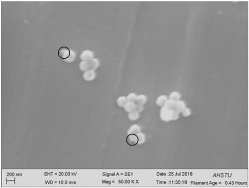
[0069] Take 10 μL streptavidin-labeled magnetic nanoparticles (Shanghai Sangong No. D110557) and 10 μL, 100 μmol / L biotin-modified CD63 aptamer (Shanghai Sangong) and mix in 0.01mol / LPBS buffer for 1 h After magnetic separation, wash twice with 0.01mol / LPBS buffer, then add 20 μL of plasma (exosomes are contained in plasma), incubate for 2 hours, and then separate with a magnet. The magnetic nanoparticle-aptamer-exosome conjugate was observed under a SEM scanning electron microscope (Carl Zeiss EVO-18) ( image 3 ), it was found that the circled part is the exosome captured by the aptamer. This shows that the CD63 aptamer provided by the present invention can specifically bind to exosomes.

Embodiment 2

[0071] 1. Preparation of nano-gold-CD63 aptamer conjugates

[0072] Take 1ml of ten-fold concentrated nano-gold (15-20nm) solution, add 10 microliters of 1mM dATP, shake with a shaker for 20min at room temperature, then add 15μl of 1% SDS at a mass concentration, shake and incubate for ten minutes, then add 50μl 0.2mol / L NaCl (the speed is controlled to add 2μl every 2-3min), then add 10μl 1OD CD63 aptamer probe, react at 60°C for 3h, centrifuge with a centrifuge (12,000rpm, 10min), remove the upper The supernatant was washed three times with PBS buffer (pH7.2-7.4), and finally the precipitate was dissolved in 1 mL nanoparticle storage solution (20 mmol / L Na 3 PO 4 12H 2 O, mass concentration 5% BSA, volume concentration 0.25% Tween20, mass concentration 10% sucrose), the conjugate solution was stored in a refrigerator at 4°C until use.

[0073] 2. Binding of biotinylated EpCam aptamer and streptavidin

[0074] 50nmol / L biotinylated EpCam aptamer

[0075] (5'-Bio-CACTACAG...


PUM

PropertyMeasurementUnit
lengthaaaaaaaaaa
widthaaaaaaaaaa

Abstract

The invention provides an aptamer group for detecting exosomes, a lateral flow aptamer biosensor and a preparation method thereof, and belongs to the technical field of exosome detection. The aptamergroup for detecting the exosomes comprises a CD63 aptamer and an EpCam aptamer, wherein the CD63 aptamer can specifically bind to the CD63 protein of the exosome, and the EpCam aptamer can specifically bind to the EPCAM protein of the exosome. The two aptamer groups are not susceptible to environmental factors such as pH and temperature and have good stability. The invention also provides a lateral flow aptamer biosensor, which is mainly based on the principle of a chromatographic test strip to spray the CD63 aptamer on a bonding pad, and spray the EpCam aptamer on a detection line, and spraya DNA probe which binds to the nanogold-labeled aptamer by base complementary pairing on a quality control line. The lateral flow aptamer biosensor can detect 500,000 exosomes and provides an innovative tool for rapid screening and diagnosis of cancer.

Description

technical field [0001] The invention belongs to the technical field of exosome detection, and in particular relates to an aptamer group for detecting exosomes, a lateral flow aptamer biosensor and a preparation method thereof. Background technique [0002] Exosomes are vesicle bodies secreted by a variety of living cells, which contain various components such as proteins and RNA. This ubiquitous nano-scale membrane structure in the body can participate in the exchange of substances and information between cells, and plays an important role in various physiological and pathological processes. Exosomes are abundant in peripheral blood, urine, saliva, ascites, amniotic fluid and other body fluids, but exosomes from different tissue sources differ in composition and function, and this difference is affected by extracellular matrix and Dynamic regulation of the microenvironment. Tumor-derived or tumor-associated exosomes are an important mechanism for regulating the occurrence ...

Claims


Application Information

Patent Timeline
Patent Type & Authority Applications(China)
IPC IPC(8): C12N15/115G01N33/53G01N33/532
CPCC12N15/115C12N2310/16G01N33/5308G01N33/532
Inventor 刘国东邱万伟刘文举钱立生张静于庆才李坤
Owner 南通拓普赛斯生物科技有限公司
Why Eureka
Try Eureka