A fluorescent biosensor for detecting dna glycosylase udg and its preparation method

A biosensor and glycosylase technology, applied in the field of biosensors, can solve the problems of long detection period, low specificity and sensitivity, and achieve the effects of fast detection speed, easy operation and short detection cycle

Active Publication Date: 2021-03-30
UNIV OF JINAN
View PDF7 Cites 0 Cited by
  • Summary
  • Abstract
  • Description
  • Claims
  • Application Information

AI Technical Summary

Problems solved by technology

[0004] In order to solve the problems of relatively low specificity and sensitivity and long detection period of the method for detecting UDG in the prior art, the present invention provides a method based on polymerase-assisted feedback rolling circle amplification with high specificity and sensitivity and fast detection speed. A biosensor for detecting DNA glycosylase UDG with endonuclease amplification fluorescence method, and also provides its preparation method

Method used


Image

  • A fluorescent biosensor for detecting dna glycosylase udg and its preparation method
  • A fluorescent biosensor for detecting dna glycosylase udg and its preparation method
  • A fluorescent biosensor for detecting dna glycosylase udg and its preparation method

Examples


Embodiment 1

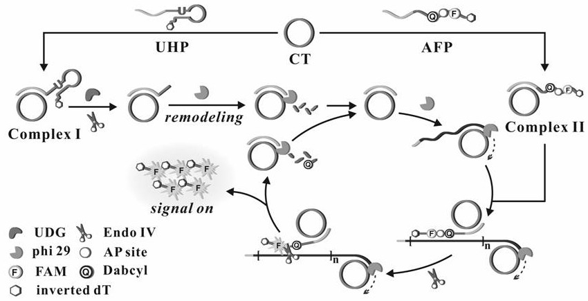
[0062] Embodiment 1 The preparation of circular template and composite probe

[0063] Prepared with 50mM Tris-HCl, 10mM MgCl 2 , T4 DNA Ligase Reaction Buffer with 10 mM DTT and 1 mM ATP. Formulated with 10mM Na 2 HPO 4 ,10mM NaH 2 PO 4 , 140mM NaCl, 1mM KCl, 1mM MgCl 2 ,1mM CaCl 2 , PBS buffer at pH=7.4.

[0064] (1) Mix 42 μL sterilized water, 6 μL linear template (10 μM), 6 μL ligation probe (10 μM) and 6 μL 10× T4 DNA ligase buffer, denature at 95°C for 5 minutes, then cool slowly to room temperature to complete hybridization , then add 3 μL T4 DNA ligase (60U / μL) to the reaction system, and react it at 16°C for 20 hours; after that, the reaction system is placed in a water bath at 65°C for 15 minutes to inactivate the T4 DNA ligation in the system enzyme.

[0065] (2) Add 3 μL of exonuclease I (20 U / μL) and 3 μL of exonuclease III (100 U / μL) to the above reaction system and react at 37 ° C for 2 h; then heat the reaction system in a water bath at 85 ° C for 10 mi...

Embodiment 2

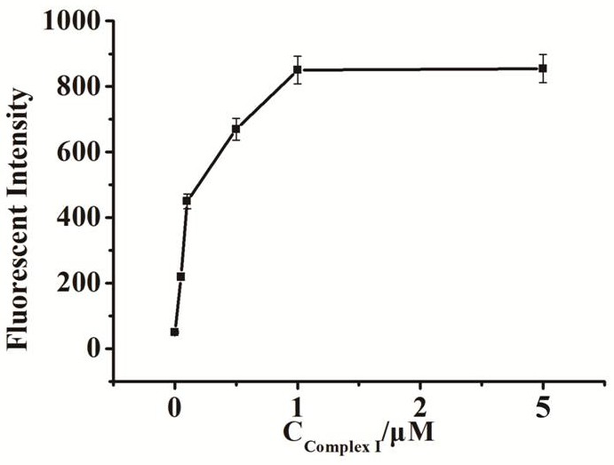
[0067] Embodiment 2 Fluorescence intensity changes with the compound probe I concentration

[0068] A method for preparing a fluorescent biosensor of the present invention, comprising the following steps:

[0069] (1) Mix 2 μL compound probe I (concentrations are 50 nM, 100 nM, 500 nM, 1 μM, 5 μM), 2 μL dNTP (1 mM), 2 μL phi29 DNA polymerase (1 U / μL), 2 μL endonuclease IV (1 U / μL) μL) in 2μL buffer (50mM Tris-HCl, 10mM MgCl 2 ,10mM (NH 4 ) 2 SO 4 , 4mM DTT, pH 7.5), add 2μL UDG enzyme solution (1U / mL) after mixing, and react at a constant temperature of 37°C for 60min after mixing;

[0070] (2) Add 2 μL of composite probe II (1 μM) to the solution in step (1), mix and react at a constant temperature of 37°C for 60 minutes;

[0071] (3) Dilute the solution obtained in step (2) with water to 100 μL, and then perform fluorescence detection; the excitation wavelength is set to 486 nm, the emission wavelength is 518 nm, and the detection range is 450 nm-530 nm, and the fluores...

Embodiment 3

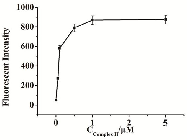
[0075] Embodiment 3 Fluorescence intensity changes with the compound probe II concentration

[0076] A method for preparing a fluorescent biosensor of the present invention, comprising the following steps:

[0077] (1) Mix 2 μL compound probe I (1 μM), 2 μL dNTP (1 mM), 2 μL phi29 DNA polymerase (1 U / μL), 2 μL endonuclease IV (1 U / μL) in 2 μL buffer (50 mM Tris-HCl ,10mM MgCl 2 ,10mM (NH 4 ) 2 SO 4 , 4mM DTT, pH 7.5), add 2μL UDG enzyme solution (1U / mL) after mixing, and react at a constant temperature of 37°C for 60min after mixing;

[0078] (2) Add 2 μL of composite probe II (concentrations are 50 nM, 100 nM, 500 nM, 1 μM, 5 μM) to the solution in step (1), mix and react at a constant temperature of 37 ° C for 60 min;

[0079] (3) Dilute the solution obtained in step (2) with water to 100 μL, and then perform fluorescence detection; the excitation wavelength is set to 486 nm, the emission wavelength is 518 nm, and the detection range is 450 nm-530 nm, and the fluorescen...


PUM


Abstract

The invention relates to the technical field of a biosensor, and particularly relates to a fluorescence biosensor for detecting UDG (Uracil-DNA Glycosylase) on the basis of a polymerase-assisted feedback rolling circle amplification and endonuclease amplification fluorescence method. The invention aims to solve problems of both low specificity and low sensitivity of a method for detecting UDG in the prior art. The biosensor for detecting the UDG on the basis of a feedback rolling circle amplification technology achieves a rolling circle amplification effect on matching of phi29 polymerase andendonuclease IV, implements fluorescence resonance energy transfer of a fluorophore and a Quenching group and performs a homogeneous reaction on mixed liquid. A preparation method comprises: constructing a circular template and a composite probe; feeding back a rolling circle amplification signal and carrying out fluorescence detection. Specific hydrolysis of the UDG on a basic group U is utilized, and by utilizing such specific reaction, the UDG can be accurately measured and meanwhile, interference can also be avoided; by utilizing endonuclease IV circle amplification, a signal amplificationeffect is achieved.

Description

technical field [0001] The invention relates to the technical field of biosensors, in particular to a fluorescent biosensor based on feedback rolling circle amplification and endonuclease signal amplification, and a preparation method thereof. Background technique [0002] DNA glycosylase UDG is an important base excision repair enzyme responsible for repairing DNA damage caused by uracil and maintaining the integrity of the genome, and the abnormal expression of UDG is also associated with various cancers. Hydrolysis of cytosine to uracil is the most common form of DNA hydrolytic damage, resulting in the conversion of G:C base pairs to A:U base pairs during DNA replication. As the initiator and "patrolman" of the base excision repair pathway, UDG has high specificity for uracil, so it can be used to recognize and catalyze the hydrolysis and splitting of N-glycosidic bonds on single-stranded or double-stranded DNA. Subsequently, the damaged base is released and creates an a...

Claims


Application Information

Patent Timeline
Patent Type & Authority Patents(China)
IPC IPC(8): G01N21/64
CPCG01N21/6402G01N21/6486
Inventor 黄加栋王敬锋刘素王玉宋晓蕾张雪王海旺赵一菡瞿晓南张儒峰
Owner UNIV OF JINAN
Why Eureka
Try Eureka