Fusion antigen for detecting echinococcosis, coding gene thereof, host cell and kit
A fusion antigen, echinococcosis technology, applied in the detection of echinococcosis fusion antigen, host cells and kits, and its coding gene field, can solve the problems of specificity and sensitivity improvement degree limitation, to improve sensitivity and specificity sexual effect
- Summary
- Abstract
- Description
- Claims
- Application Information
AI Technical Summary
Problems solved by technology
Method used
Image

Examples
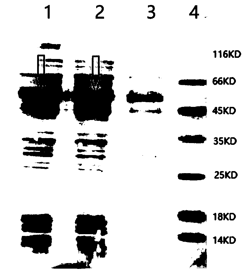
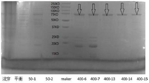
Embodiment 1
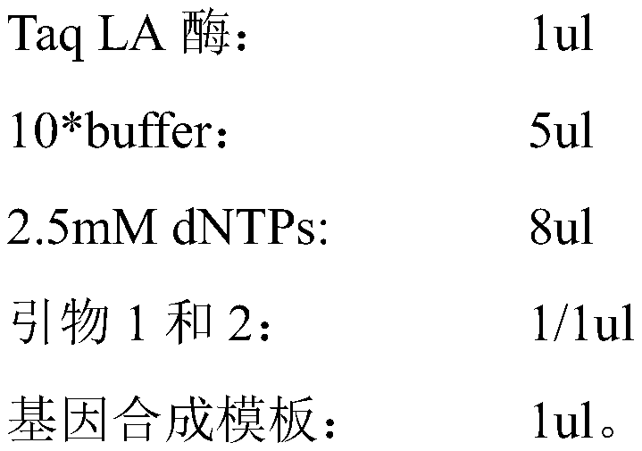
[0048] This example is a preparation example of the fusion antigen.
[0049] 1. Synthesize the gene of the fusion antigen, its sequence is shown in SEQ ID NO:6. Amplify the reaction using the following reaction system:
[0050] 50ul system
[0051]
[0052] in:
[0053] The sequence of primer 1 is: CGCGGATCCATGAAAGATGAGCCAAAAGCA, which contains a BamHI restriction site;
[0054] The sequence of primer 2 is: CCCTCGAGTTATTTGAGGTTGGCCAGCT, which contains an XhoI restriction site.
[0055] The PCR amplification conditions are:
[0056] 35 cycles of reaction process:
[0057] 94°C: 7min
[0058] 94°C: 30s
[0059] 60°C: 30s
[0060] 72°C: 2min
[0061] 72°C: 10min
[0062] 4°C: for eve.
[0063] 2. Identification by double enzyme digestion
[0064] The reaction system and conditions for double enzyme digestion identification are as follows:
[0065] System 40ul:
[0066]
[0067] React at 37°C for two hours.
Embodiment 2
[0156] This embodiment is an example of the preparation and use of the reagent strip. Specifically, the detection area (T line) on the NC membrane of the test card is coated with the echinococcosis antigen of Example 1, and the quality control area (C line) is coated with anti-hydatid antibody.
[0157] (1) Coating: Dilute the fusion antigen of Example 1 to 0.2 mg / ml as a T-line coating solution, and dilute the anti-hydatid antibody to 2 mg / ml as a C-line coating solution. Coat the T-line coating solution and the C-line coating solution on the nitrocellulose membrane through a film spraying machine. to dry.
[0158] (2) UCP-labeled antigen: The UCP-labeled antigen in this kit is obtained through the following steps:
[0159] a) Weigh 10 mg of UCP particles and place them in a conical flask;
[0160] b) Add 10ml pH=7.2 0.20M PB;
[0161] c) Add 0.5 mg of the antigen of Example 1 to the UCP particle suspension, then add anhydrous glutaraldehyde to a final concentration of 1%...
Embodiment 3
[0176] This embodiment is an application example of the reagent strip. During the test, the sample is added to the sampling point of the test card, and the liquid is chromatographed under the capillary effect. The substance will be bound by the antigen coated on the T line, and a solid-phase UCP particle-echinococcosis marker antigen-echinococcosis antibody-echinococcosis antigen complex will be formed at the position of the T line, and a solid-phase complex will be formed at the C line Anti-hydatid antibody-labeled antigen-UCP complex. Finally, the detection result is obtained by detecting the emitted light signal of the upconverted luminescent particle.
[0177] Table 1 - Echinococcosis serum antibody detection results
[0178]
[0179] Comparative detection of echinococcosis serum antibody detection reagents and commercially available reagents:
[0180] Comprehensive evaluation of 108 cases of echinococcosis positive samples (ELISA) and 162 cases of normal human serum...
PUM
| Property | Measurement | Unit |
|---|---|---|
| Sensitivity | aaaaa | aaaaa |
Abstract
Description
Claims
Application Information
 - R&D
- Intellectual Property
- Life Sciences
- Materials
- Tech Scout
- Unparalleled Data Quality
- Higher Quality Content
- 60% Fewer Hallucinations
Browse by: Latest US Patents, China's latest patents, Technical Efficacy Thesaurus, Application Domain, Technology Topic, Popular Technical Reports.
© 2025 PatSnap. All rights reserved.Legal|Privacy policy|Modern Slavery Act Transparency Statement|Sitemap|About US| Contact US: help@patsnap.com