
A method for preparing thyroid and breast fine-needle aspiration cell tissue block

A technology of cell tissue and thyroid, which is applied in the field of preparation of thyroid and breast fine-needle aspiration cell tissue blocks, which can solve the problems of inappropriate fine-needle aspiration mammary gland specimen cell smears and difficult accurate diagnosis, achieving good application effect and saving detection costs , good preservation effect

Active Publication Date: 2021-08-10
马晓丽
View PDF6 Cites 0 Cited by
  • Summary
  • Abstract
  • Description
  • Claims
  • Application Information

AI Technical Summary

Problems solved by technology

[0006] The purpose of the present invention is to provide a method for preparing thyroid and breast fine-needle aspiration cell tissue blocks, so as to solve the problem that the existing tissue block preparation methods are not suitable for fine-needle aspiration of thyroid and breast specimens and cell smears, which are difficult to diagnose accurately

Method used


Image

  • A method for preparing thyroid and breast fine-needle aspiration cell tissue block
  • A method for preparing thyroid and breast fine-needle aspiration cell tissue block
  • A method for preparing thyroid and breast fine-needle aspiration cell tissue block

Examples


Embodiment 1

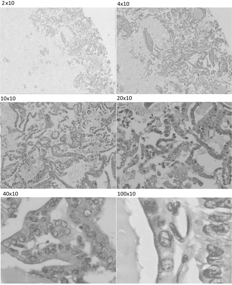
[0025] Example 1: Preparation of Thyroid Fine Needle Aspiration Cell Tissue Block.

[0026] 1. Pre-experimental steps: Take non-infectious pleural effusion and centrifuge twice to obtain 100mL of supernatant, respectively take 0.5mL, 1mL, 1.5mL, and 2mL of supernatant and inject them into disposable 10mL centrifuge tubes, add 95% of the same volume Gently mix with ethanol, centrifuge at 2000 r / min for 10 min, observe the size of the precipitated mass, and take the mass at the bottom of the centrifuge tube with a height of about 0.3cm (volume is about 1.0cm*1.0cm*0.3cm) It is used as a sample for backup, and the lumps of other centrifuge tubes are discarded.

[0027] Discard the supernatant of the selected sample, and add 5-10 times of 4% neutral buffered formaldehyde fixative. After solidification, the solidified cell pellets in the centrifuge tube were taken out and put into embedding paper, and then put into a dehydrated embedding box. Paraffin tissue specimens were process...

Embodiment 2

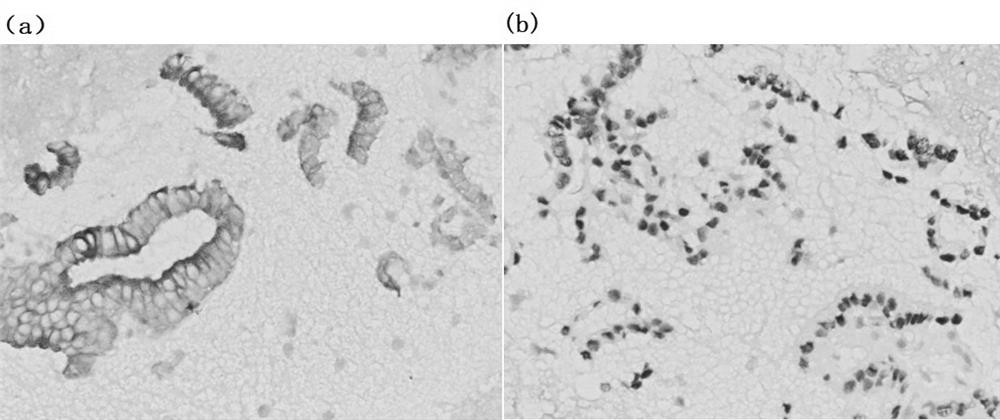
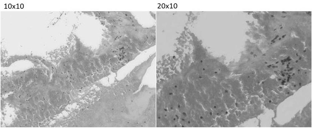
[0032] Example 2: Preparation of mammary fine needle aspiration cell tissue block.

[0033] 1. Pre-experimental steps: Take non-infectious ascites and centrifuge twice to obtain 100mL of supernatant, respectively take 0.5mL, 1mL, 1.5mL, and 2mL of supernatant and inject them into disposable 10mL centrifuge tubes, add 95% of the same volume Gently mix with ethanol, centrifuge at 2000 r / min for 5 minutes, observe the size of the precipitated mass, and take the mass at the bottom of the centrifuge tube with a height of about 0.3cm (volume is about 1.0cm*1.0cm*0.3cm) It is used as a sample for backup, and the lumps of other centrifuge tubes are discarded.

[0034] Discard the supernatant of the selected sample, and add 5-10 times of 4% neutral buffered formaldehyde fixative. After solidification, the solidified cell pellets in the centrifuge tube were taken out and put into embedding paper, and then put into a dehydrated embedding box. Paraffin tissue specimens were processed by ...


PUM


Abstract

The invention provides a method for preparing thyroid and mammary gland fine needle aspiration cell tissue block. It includes the following steps: a. Centrifuge non-infectious pleural effusion or ascites in advance to obtain pleural effusion or ascites supernatant; b. Take pleural effusion or ascites supernatant and inject it into a centrifuge tube; c. Thyroid or mammary gland punctured with a fine needle Inject the sample into the centrifuge tube, then immediately take 95% ethanol and inject it into the centrifuge tube; d, centrifuge the centrifuge tube and discard the supernatant to obtain the cell sediment, then add 4% neutral buffered formaldehyde fixative solution, and the cells in the centrifuge tube After the precipitate had solidified, routine embedding was performed using paraffin tissue specimen processing procedures. The preparation method of the invention can ensure no loss of tissue fragments and free-floating single cells, and the shape and structure of the cytoplasm are well preserved.

Description

technical field [0001] The invention relates to the technical field of cell tissue block preparation, in particular to a method for preparing thyroid and mammary gland cell tissue block by fine needle aspiration. Background technique [0002] The incidence of thyroid cancer and breast cancer worldwide is increasing rapidly year by year, among which the incidence of thyroid cancer is increasing at a rate of 6% per year. At present, the average incidence rate of thyroid cancer in China is 7.7 / 100,000, ranking fifth in the incidence rate of malignant tumors. 85% of thyroid adenocarcinomas are papillary carcinoma (Papillary Thyroid Carcinoma, PTC). A special biological behavior of PTC is the early occurrence of regional lymph node metastasis. Although the prognosis of differentiated thyroid cancer is good, about 30% of thyroid cancer patients will relapse during nearly 30 years of follow-up, and a small number of patients will eventually die of thyroid cancer. The preoperativ...

Claims


Application Information

Patent Timeline
Patent Type & Authority Patents(China)
IPC IPC(8): G01N1/28G01N1/30G01N1/36
CPCG01N1/30G01N1/36
Inventor 马晓丽
Owner 马晓丽
Learn More