Marker (MYC) for predicting cervical lesions occurrence of HPV positive patients
A technology of cervical lesions and biomarkers, applied in the application field of tumor markers in predicting the prognosis of cervical cancer, can solve the problems of low sensitivity, low specificity and sensitivity, missed diagnosis and missed treatment of patients, etc., and achieve a false positive rate Low, sensitive diagnosis or, the effect of reducing the workload
- Summary
- Abstract
- Description
- Claims
- Application Information
AI Technical Summary
Problems solved by technology
Method used
Image
Examples
Embodiment 1
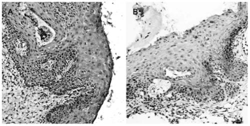
[0022] Example 1: Using immunohistochemical staining to detect the expression of MYC protein in different tissue samples
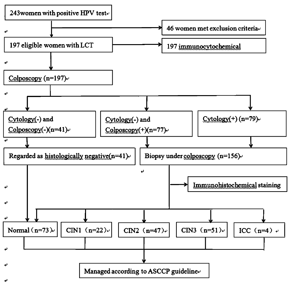
[0023] Cases: From September 2015 to September 2017, HPV-positive patients who underwent cervical cancer screening at the First Affiliated Hospital of Sun Yat-sen University. Patients were tested for both HPV and LCT, or they had HPV test results from another authoritative medical institution. A total of 243 HPV-positive women were recruited, and samples from 197 eligible subjects who agreed to undergo colposcopy were collected for the study.
[0024] Exclusion criteria: (1) patients with a history of CIN or cervical cancer treatment; (2) patients younger than 30 years old or older than 65 years old; (3) patients who were pregnant or breastfeeding.
[0025] Among the 197 patients who underwent colposcopy, 73 cases were normal, 22 cases were CIN1, 47 cases were CIN2, 51 cases were CIN3, and 4 cases were ICC (the operation process was as follows: figure 1 ...
Embodiment 2
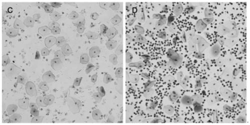
[0040] Example 2. Immunohistochemical method to detect the expression of MYC protein in different cell samples.
[0041] (1) Dewaxing and hydration: put the prepared cell smear in a 60°C incubator and bake for 2 hours, then immerse in xylene 3 times, 5 minutes each time; 2 times in absolute ethanol, 5 minutes each time; In 95%, 85%, and 75% ethanol for 5 minutes each; wash with PBS 3 times, 5 minutes each time.
[0042] (2) Eliminate endogenous peroxidase: Remove the liquid around the tissue on the smear, place it flat in a wet box, add 3% hydrogen peroxide solution dropwise, incubate at room temperature for 10-15 minutes, wash with PBS 3 times, 5 minutes each time .
[0043] (3) Antigen retrieval: Put the smear into citrate buffer, put it into a pressure cooker and heat it to boiling, 800w, cover the pressure valve and wait for 1-4 minutes after spraying. The temperature of the buffer was naturally cooled to room temperature, and washed 3 times with PBS (containing Triton X...
Embodiment 3
[0053] Example 3 Verifies the correlation between MYC gene and human papillomavirus positive patients
[0054] The corresponding MYC protein expression levels in different stages of cervical lesions (normal, CIN1, CIN2, CIN3 and invasive cancer) in the aforementioned cases of cervical lesions were analyzed to obtain the frequency and proportion of MYC staining intensity at different stages. The results are shown in Table 1:
[0055] Table 1: Frequency and proportion of staining intensity of MYC in different stages of cervical lesions
[0056]
[0057]
[0058] CIN = cervical intraepithelial neoplasia; ICC = invasive cervical cancer
[0059] It can be seen from Table 1 that MYC staining was positive in CIN2+ tissues and negative in CIN1- tissues, indicating that the trend of MYC expression intensity was positively correlated with the severity of cervical disease.
PUM
 Abstract
Description
Claims
Application Information
 - R&D
- Intellectual Property
- Life Sciences
- Materials
- Tech Scout
- Unparalleled Data Quality
- Higher Quality Content
- 60% Fewer Hallucinations
Browse by: Latest US Patents, China's latest patents, Technical Efficacy Thesaurus, Application Domain, Technology Topic, Popular Technical Reports.
© 2025 PatSnap. All rights reserved.Legal|Privacy policy|Modern Slavery Act Transparency Statement|Sitemap|About US| Contact US: help@patsnap.com


