Device and method for in-vivo micro-bubble control and imaging

A microbubble and imaging technology, which is applied to the human tubular structure device, prosthesis, ultrasonic therapy, etc., can solve the problems of increased risk of systemic hemorrhage, difficulty in ultrasonic imaging, lumen stenosis, etc., to improve drug-carrying efficiency and reduce production. cost, the effect of reducing toxic and side effects

Active Publication Date: 2019-10-18
SHANDONG UNIV QILU HOSPITAL +1
View PDF6 Cites 6 Cited by
  • Summary
  • Abstract
  • Description
  • Claims
  • Application Information

AI Technical Summary

Problems solved by technology

For example, arterial stenosis caused by atherosclerosis, although the use of intravascular stents can effectively solve the problem of stenosis of the lumen, but because atherosclerosis cannot be cured only by the support of stents, it is accompanied by stent implantation. In-stent restenosis makes maintaining stent patency a new problem and challenge
[0004] Although there is currently a drug-eluting stent technology to slow down in-stent restenosis, this technology has caused new problems: the eluted drug is an anticancer drug such as sirolimus, which leads to delayed healing of the va

Method used


Image

  • Device and method for in-vivo micro-bubble control and imaging
  • Device and method for in-vivo micro-bubble control and imaging
  • Device and method for in-vivo micro-bubble control and imaging

Examples


Example Embodiment

[0045] Embodiment one

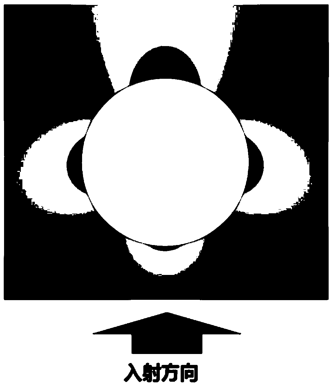
[0046] The sound wave manipulation of particles mainly uses the particles in the sound field to produce scattering, reflection, refraction, absorption and other effects on the sound waves, resulting in the exchange of momentum carried by the sound field between the sound field and the particles, and the movement of the particles is captured by the force. For bubbles and rigid particles of the same size, the acoustic scattering cross-sectional area of ​​the bubble resonance can reach hundreds of millions of times the acoustic scattering cross-sectional area of ​​the rigid particle, so the acoustic radiation force of the bubble in the sound field is much higher than that of the rigid particle. Force, so the bubbles are easier to manipulate. And because of the excellent scattering properties of bubbles, both linear and nonlinear components of the scattering signal can be used for imaging to improve image quality.

[0047] Therefore, the present invention ...

Example Embodiment

[0078] Embodiment two

[0079] In one or more embodiments, a method for in vivo manipulation of microvesicles is disclosed, comprising:

[0080] Calculate the delay time for manipulating each array element of the transducer according to the spatial position of the area of ​​interest, generate an excitation signal for manipulating the transducer according to the delay time, and stimulate the corresponding array elements to emit sound waves, and the sound waves emitted by each array element propagate to the area of ​​interest Regionally and focused excitation of stent-shaped implanted structures in vivo;

[0081] The stent-type in vivo implant structure generates a local acoustic field through resonance, and the drug-loaded microbubbles are captured and aggregated by the stent-type in vivo implant structure under the action of the local sound field, and the microbubbles are broken by changing the ultrasonic intensity to release drug particles.

Example Embodiment

[0082] Embodiment Three

[0083] In one or more embodiments, a method for imaging microvesicles in vivo is disclosed, comprising:

[0084] Calculate and manipulate the delay time of each element of the transducer according to the spatial position of the region of interest, generate the excitation signal of the imaging transducer according to the delay time, excite the corresponding array element to emit sound waves, synthesize the focal point in the space, and control each element in the space point to scan;

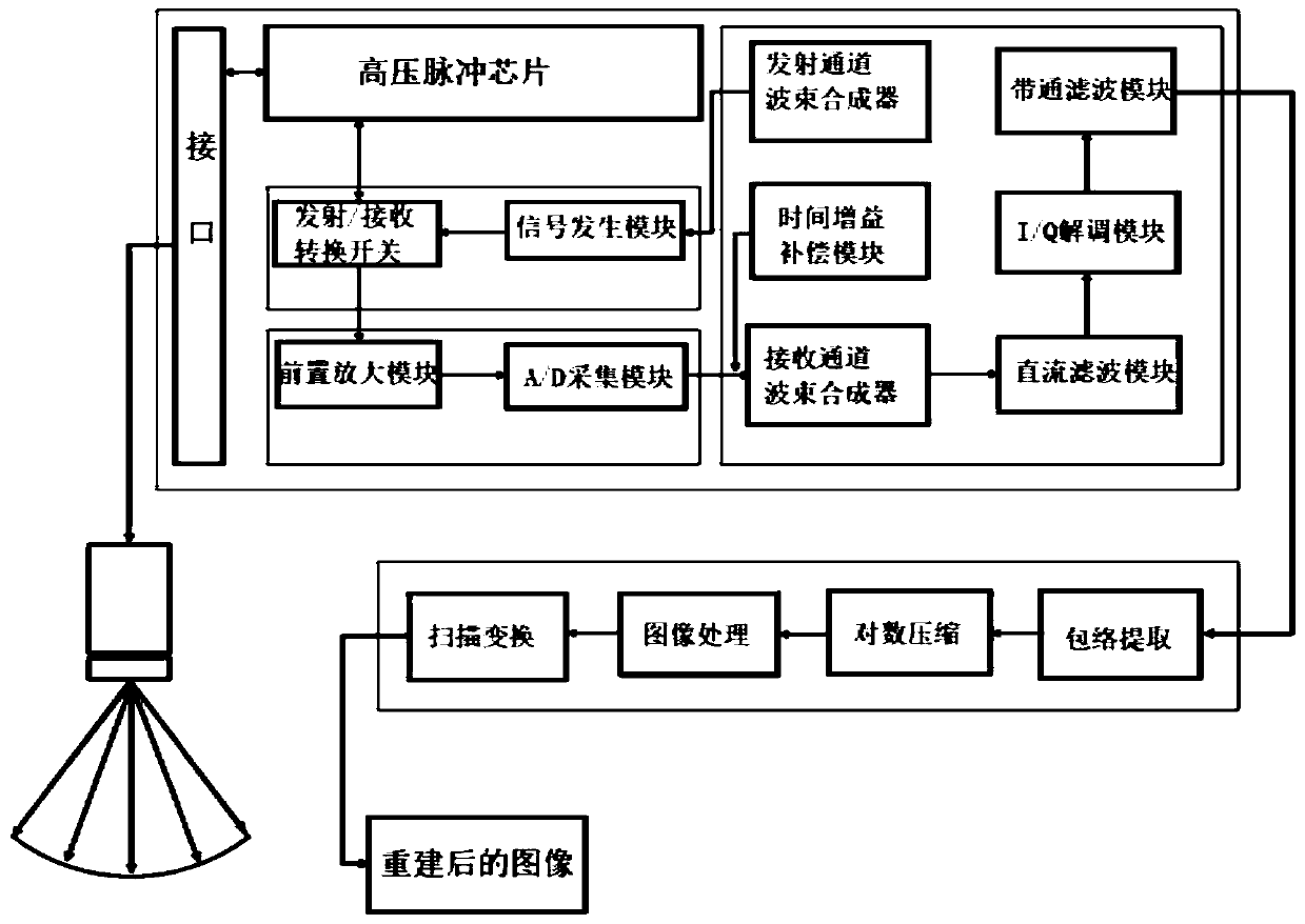
[0085] Switch from transmitting mode to receiving mode, receive echo signals from different positions, and the echo signals are sequentially pre-amplified, A / D acquired and time gain compensated; the echo signals are beam-formed according to the delay time of different spatial points ;

[0086] The echo signal after beamforming is subjected to DC filtering, I / Q demodulation and band-pass filtering in sequence, and the fundamental wave component and harmonic component a...


PUM


Abstract

The invention discloses a device and method for in-vivo micro-bubble control and imaging. The device comprises a support type in-vivo implanting structure, drug-loading micro-bubbles, a control arraytransducer unit and an imaging array transducer unit. Drug particles are loaded on the surfaces of the micro-bubbles or in the micro-bubbles, the drug-loading micro-bubbles are captured and gathered by the support type in-vivo implanting structure under the action of a local-area sound field, the micro-bubbles are broken by changing the ultrasonic intensity, and the drug particles are released. The control array transducer unit is used for transmitting power ultrasonic to excite the support type in-vivo implanting structure to vibrate and capture the drug-loading micro-bubbles in blood vessels. The imaging array transducer unit is used for emitting pulse waves, receiving echo signals from different positions, and imaging the micro-bubbles, the support type in-vivo implanting structure andtissue structures. According to the device, the resonance sound field generated by the support type in-vivo implanting structure is utilized, so that the micro-bubble aggregation in the blood vesselsis controlled; an ultrasonic transducer used for imaging is additionally arranged, and the micro-bubble control effect can be observed in real time.

Description

technical field [0001] The invention belongs to the technical field of ultrasonic drug delivery, and in particular relates to a device and method for manipulation and imaging of microbubbles in vivo. Background technique [0002] The statements in this section merely provide background information related to the present invention and do not necessarily constitute prior art. [0003] For the treatment of diseases, it is often necessary to implant artificial prostheses in the human body, such as artificial skulls, artificial joints, artificial bones, and various intracavitary implants, such as vascular stents, tracheal stents, esophageal stents, urethral stents, etc. For many diseases, these implants cannot cure the primary disease. In order to continuously intervene in the disease process or maintain the patency of the implant in the cavity, it is necessary to continue to deliver drugs that interfere with the disease process to the implant in subsequent treatments. the impla...

Claims


Application Information

Patent Timeline
IPC IPC(8): A61N7/00A61F2/07
CPCA61F2/07A61F2250/0067A61N7/00A61N2007/0039A61N2007/0043
Inventor 张鹏飞李飞苏敏毛洋
Owner SHANDONG UNIV QILU HOSPITAL
Why Eureka
Try Eureka