

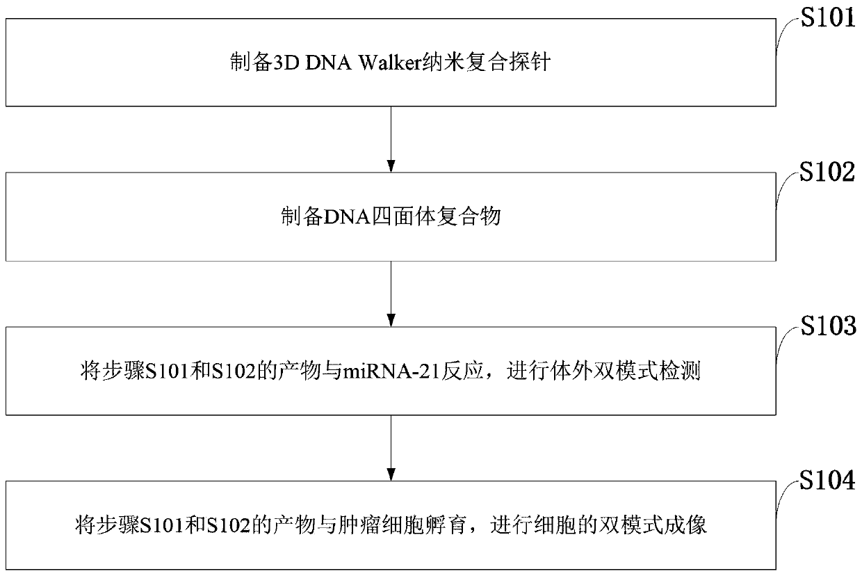
Method for detecting tumor cell marker miRNA-21 and tumor cells
A technology of miRNA-21 and tumor cells, which is applied in the detection field of tumor cell marker miRNA-21 and tumor cells, can solve the problems of low signal, high accuracy, and limited sensitivity of detection results, and achieve the detection of fewer samples required , Large linear range and high sensitivity
- Summary
- Abstract
- Description
- Claims
- Application Information
AI Technical Summary
Problems solved by technology
Method used
Image

Examples
Embodiment 1
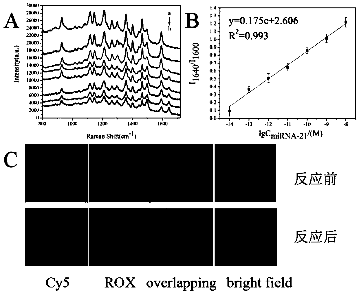
[0078] Raman assay was performed on different concentrations of miRNA-21, image 3 -A is the Raman signal of different concentrations of miRNA-21, from curve a to curve h are the corresponding Raman signal curves of 0nM, 10nM, 1nM, 100pM, 10pM, 1pM, 0.1pM, 0.01pM concentrations of miRNA-21 picture. Choose 1600cm -1 It is the Raman characteristic absorption peak of Cy5, 1640cm -1 is the Raman characteristic absorption peak of Rox, the peak intensity of its peak position is respectively subtracted from the blank processing, and using I 1640 / I 1600 Performing a linear analysis for the final linear analysis object yields image 3 -B.
Embodiment 2
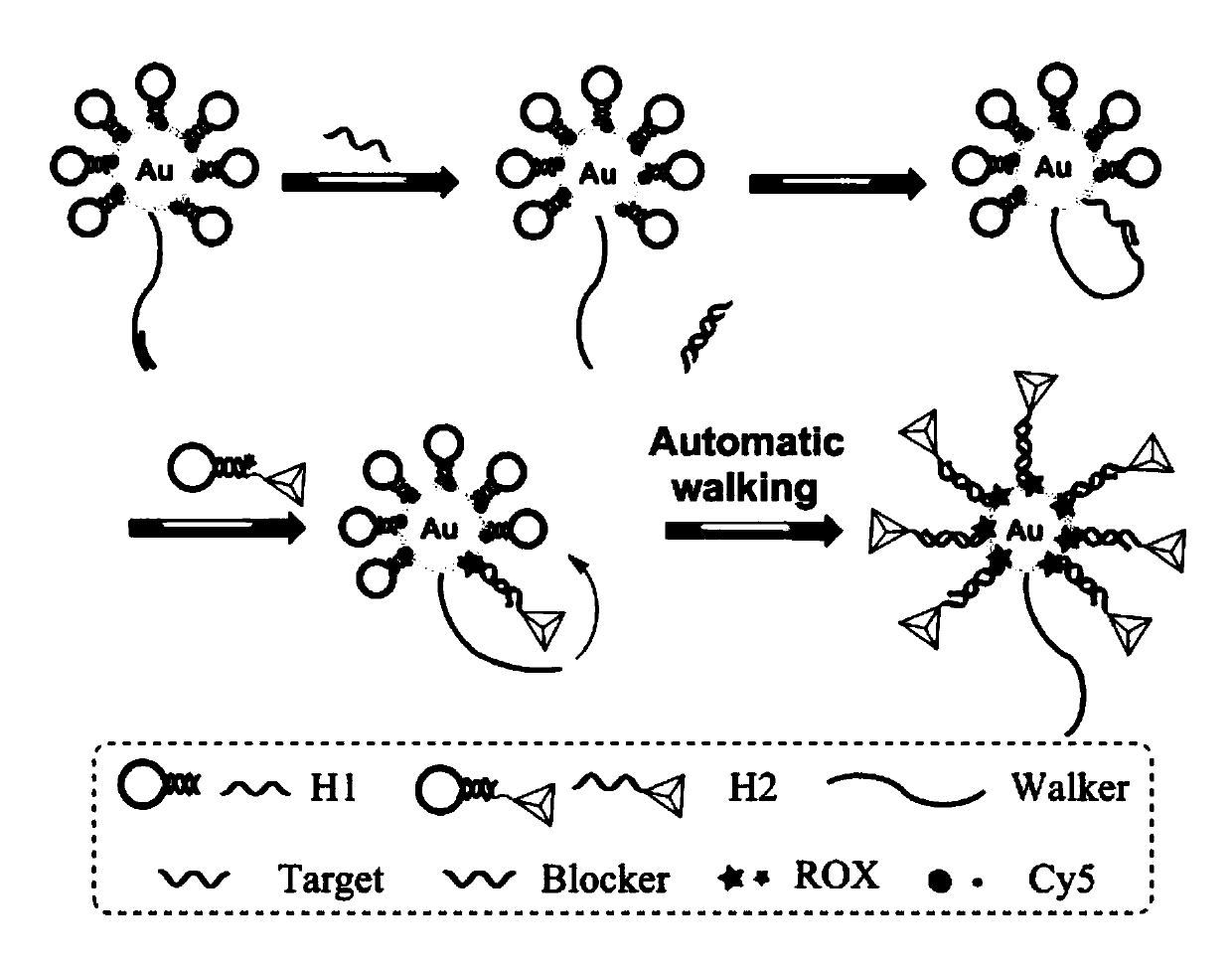
[0080] For Raman imaging detection of tumor cells such as image 3 As shown in -C, 200 μL of trypsinized Hela cells were seeded on a gold glass slide overnight, and 20 μL of biological barcode probe and 20 μL of 1 μM hairpin DNA H were added. 2 -DNA tetrahedral complexes, incubated for different times. Fix the cell culture dish on the microscope stage, and perform SERS imaging in the state of cell culture. Use the 633nm laser of the Raman spectrometer and perform SERS cell imaging with a 50x objective lens. Depend on image 3 In -C, it can be seen that the Raman signal intensity of Cy5 was significantly weakened after the reaction, and the Raman signal intensity of Rox was significantly enhanced, indicating the presence of miRNA-21 in Hela cells.
Embodiment 3
[0082] Fluorescent assays were performed on different concentrations of miRNA-21, Figure 4 -A is the fluorescence signal of Cy5 of different concentrations of miRNA-21, Figure 4 -B is the fluorescence signal of Rox with different concentrations of miRNA-21, from curve a to curve h are the fluorescence signal curves corresponding to the concentrations of miRNA-21 of 0nM, 100nM, 50nM, 10nM, 5nM, 1nM, 0.5nM, 0.1nM respectively picture. Fluorescence spectra were determined for Cy5 using an excitation wavelength of 648 nm with a maximum emission wavelength of 688 nm. Fluorescence spectra were determined for Rox using an excitation wavelength of 550 nm with a maximum emission wavelength of 610 nm. The peak intensity was deducted from the blank respectively, and the F 610 / F 688 Linear analysis is performed for the final linear analysis object to get Figure 4 -C.
PUM
 Abstract
Description
Claims
Application Information