Lactobacillus casei SYF-08 capable of adsorbing heavy metal lead and application of lactobacillus casei SYF-08
A technology of SYF-08, Lactobacillus casei, applied in Lactobacillus, application, bacteria used in food preparation, etc., can solve the problem of no reported separation, reduce lead absorption and chronic lead poisoning, stabilize content, reduce recognition effects of functional impairment
Pending Publication Date: 2019-12-31
GUANGDONG HUANKAI BIOLOGICAL SCI & TECH CO LTD +1
2 Cites 3 Cited by
AI-Extracted Technical Summary
Problems solved by technology
However, these strains are not isolated from plants, and there is no report...
Abstract
The invention discloses lactobacillus casei SYF-08 capable of adsorbing heavy metal lead and an application of the lactobacillus casei SYF-08. The lactobacillus casei SYF-08 is separated from excrement and urine of healthy newborns, has the capacity of bearing lead ions and highly adsorbing the lead ions, can be stably colonized in intestinal tracts and can adsorb the lead ions entering the intestinal tracts. The invention also discloses an application of bacterial liquid of the strain SYF-08 to preparation of food additives, foods or medicines having the efficacy of preventing and/or assisting in treating chronic lead poisoning.
Application Domain
Antibacterial agentsBacteria +4
Technology Topic
ChemistryUrine +6
Image
Examples
- Experimental program(5)
Example Embodiment
[0028] Example 1
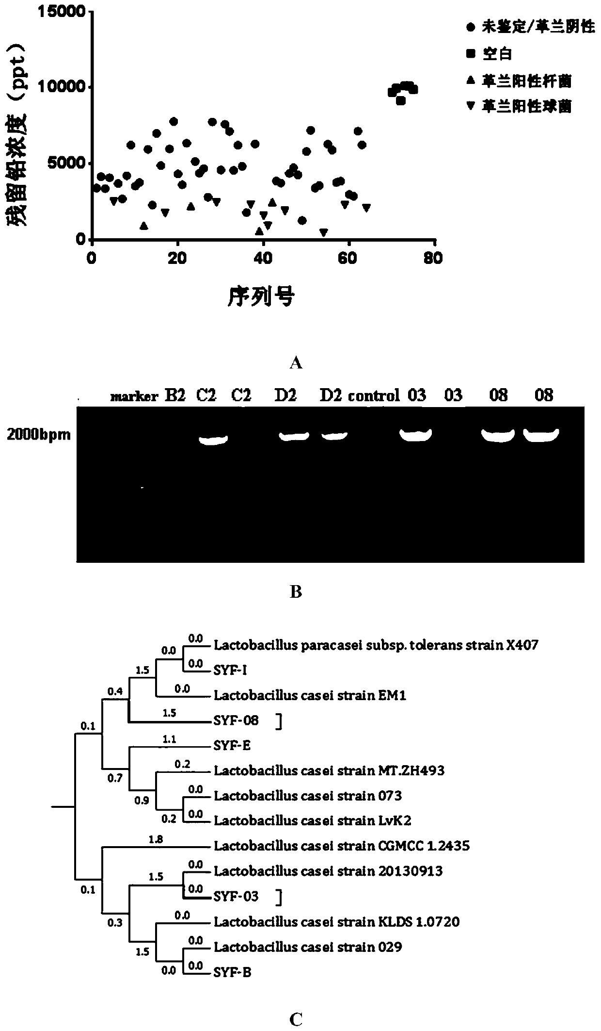
[0029] Isolation of Lactobacillus casei strains
[0030] Take an appropriate amount of fresh feces from healthy newborns who are exclusively breastfed, and dilute it with a 5mg/L lead solution at a solid-to-liquid ratio of 1:1000 (g/ml). Take 5μl of sample and inoculate it in modified MRS medium (central diffusion method) (scribing plate radially from the center to the edge). Add 200μl of lead solution with a concentration of 10000mg/L dropwise to the center of the plate, and anaerobic culture at 37℃ for 48h. Pick a single colony close to the center of the agar plate and inoculate it into MRS liquid medium for enrichment culture.
[0031] After culturing for 24 hours, dilute with sterile PBS to prepare a bacterial suspension with a wavelength of 600nm and OD=1; centrifuge (8000rpm, 5min), remove the supernatant, and add 1ml of 10ppm lead acetate solution respectively; mix well and stand at 37℃ for 1h Centrifuge (12000rpm, 5min) to take the supernatant and dilute, use inductively coupled plasma mass spectrometer (ICP-MS) to detect the supernatant lead concentration ( figure 1 A).
[0032] The modified MRS medium formula is: peptone 10g, yeast extract 5g, beef extract 10g, glucose 20g, sodium acetate 5g, diammonium citrate 2g, Tween 80 1mL, magnesium sulfate 0.58g, manganese sulfate 0.05g, phosphoric acid Dipotassium hydrogen 2g, agar 15-17g, water 1000Ml, pH adjusted to 6.12~6.2.
[0033] Select strains with a lead absorption rate of ≥75% to continue to culture, Gram stain, and screen out Gram-negative, spores or flagella strains, then perform molecular identification and traditional biochemical identification.
[0034] The total bacterial DNA was extracted with the TIANamp Bacteria DNA Kit, and the extraction method steps were performed according to the kit instructions. The extracted DNA was amplified by PCR using universal primers for lactic acid bacteria 16S rDNA.
[0035] The nucleotide sequence of the universal primer pair for 16S rDNA is:
[0036] Forward primer 27f: 5'-AGAGTTTGATCCTGGCTCAG-3' (SEQ ID NO.1),
[0037] Reverse primer 1492r: 5'-GGTTACCTTGTTACGACTT-3' (SEQ ID NO. 2);
[0038] The primers were synthesized by Shanghai Shenggong Biological Co., Ltd.
[0039] A total of 20μl PCR amplification reaction system, 2μl template, 10μl TaKaRa Premix TaqTM, 1μl each of the forward primer and reverse primer, dd H 2 O 6μl. In the negative control reaction system, the template is dd H 2 O instead, the other ingredients are the same.
[0040] PCR amplification reaction conditions: 94℃5min; 94℃60s, 60℃60s, 72℃90s, 30 cycles; 72℃10 min; 4℃ storage.
[0041] Result: Observe the position of the target band, B2, C2, D2, and 03 are candidate strains screened in the same batch, 08 is Lactobacillus casei SYF-08 (see figure 1 B). (The samples of C2, D2, 03, and 08 in the figure are repeated once for each experiment)
[0042] After PCR electrophoresis, the gel was cut and the target band gel was recovered and sequenced by Shanghai Shenggong Biological Co., Ltd. In the NCBI database of the United States, BLAST software tools were used to align the 16S rDNA gene sequences of Lactobacillus casei, and MEGA7.0 was used to construct a phylogenetic tree.
[0043] The results showed that the sequencing results of Lactobacillus casei SYF-08 showed 98.5% homology with the 16S rDNA sequence of Lactobacillus casei. The SYF-08 strain was identified as Lactobacillus casei and named SYF-08, which was obtained by screening in this example Lactobacillus casei. (see figure 1 C).
[0044] The Lactobacillus casei SYF-08, which is screened in this example and has a strong ability to adsorb lead ions, was deposited in the Guangdong Provincial Microbial Culture Collection on September 3, 2018. The classification is named Lactobacillus casei, and the deposit number is GDMCC No: 60441.
[0045] As shown in Table 1, the following biochemical metabolites of the candidate strain SYF-08 were detected and isolated according to the instructions of the new microbial trace biochemical series identification tube: esculin, mannitol, salicin, sorbitol, raffinose, Maltose, sucrose, cellobiose, hippurate, rhamnose, arabinose, xylose, galactose, nitrite, catalase, hydrogen sulfide.
[0046] Results: Table 1 shows that the physiological and biochemical characteristics of this strain are basically consistent with those of general Lactobacillus casei.
[0047] Table 1 Biochemical identification results of Lactobacillus casei STF-08
[0048]
[0049]
[0050] Determination of in vitro lead adsorption capacity of Lactobacillus casei SYF-08 strain
[0051] Take Lactobacillus casei SYF-08 bacterial liquid, centrifuge to remove the supernatant, and concentrate the bacterial cells. Dilute it to lead water of various concentrations at a mass ratio of 0.1g/L. Mix well, let it stand at 37°C for 1 hour and centrifuge (12000rpm, 5min), take the supernatant and dilute, and detect the lead concentration by ICP-MS. ICP-MS detection and analysis conditions are: wavelength 220.353nm, plasma flow 15L/min, atomizing gas flow rate 0.8L/min, auxiliary gas flow rate 0.2L/min, RF power 1100w, plasma observation direction is axial, sample flow rate 1.5mL/min.
[0052] Set up 10 parallel controls for each group to calculate the adsorption rate.
[0053] Lead adsorption rate = (original lead liquid concentration-sample lead concentration) / original lead liquid concentration × 100%
[0054] The results show that when the lead concentration is 10ppm and 250ppm, the adsorption rate of Lactobacillus casei SYF-08 is 83.15±3.17% and 51.00±6.81%, respectively, so SYF-08 has lead adsorption capacity. (see figure 2 A).
[0055] Take the Lactobacillus casei SYF-08 bacterial liquid, concentrate the bacterial cell by centrifugation, and dilute the bacterial suspension with sterile PBS. LoVo cells are resuscitated, and the number of inoculated cells is about 10 after stable 5 Put each cell into a 24-well plate. When the cell length is a single layer and cover the bottom of the well, wash 3 times with sterile PBS. Add 500μl of RPMI-1640 basal medium (C11875500BT) to each well. Grouping and treatment are as follows:
[0056] Positive control (positive control): add lead solution, culture for 3h;
[0057] Competition group (competition): the corresponding concentration of lead solution +10 6 CFU SYF-08 was cultured for 3 hours;
[0058] Exclution: 10 6 After CFU SYF-08 is incubated for 1.5 hours, add the corresponding lead solution and continue to incubate for 3 hours;
[0059] Substitution group (substitution): After adding the corresponding concentration of lead solution and incubating for 1.5 hours, add 10 6 CFU SYF-08 continued to be cultured for 1.5 hours.
[0060] Set two lead concentrations of 50ppm and 100ppm, and each group has 3 multiple holes. Aspirate the supernatant, centrifuge (12000rpm, 5min), dilute, and detect the lead concentration of the supernatant. Add 500μl of sterile deionized water to each well, let stand for 10 minutes, blow the bottom of the hole, aspirate the solution, centrifuge (12000rpm, 5min), dilute, and detect the lead concentration in the cells (see figure 2 B and 2C).
[0061] The SYF-08 strain was detected by the same experimental operation.
[0062] Table 2 Comparison of lead adsorption rate between SYF-08 and SYF-B
[0063] Strain 10ppm 50ppm 250ppm SYF-B 85.22% 86.27% 36.71% SYF08 83.15% 85.15% 51.00%
[0064] Results: Table 2 shows that there is no significant difference in the adsorption capacity of SYF-B and SYF08 in low-lead solutions, while SYF08 is more stable when the lead solution concentration is 250ppm, and the adsorption performance is better than SYF-B, and it has more application prospects.
Example Embodiment
[0065] Example 2 Test of probiotic properties of Lactobacillus casei SYF-08
[0066] 1. Acid and bile resistance
[0067] Prepare artificial gastric juice with pH values of 2.0, 3.0, 4.0. Take Lactobacillus casei SYF-08 and dilute to 10 with sterile PBS 9 CFU/ml bacterial suspension. Pipet 10μl of bacterial suspension in 990μl of artificial gastric juice with different pH values and incubate anaerobicly at 37°C for 1 to 3 hours. Use the dilution drop plate method to determine the number of viable cells at the beginning and after the culture.
[0068] Bile resistance test: bile salt comprehensive test (bile salt concentration 0.3%) test strains that have passed acid resistance screening. The counted strains were placed in pH 3.0 phosphate buffer, incubated anaerobicly at 37°C for 2 hours, and 10 μL was inoculated into 990 μL of a 0.3% bile salt solution, incubated anaerobic at 37°C for 8 hours, and the original and The number of viable bacteria after culture (see Table 3).
[0069] The SYF-08 strain was detected by the same operation. (The operation is the same as above)
[0070] Results: Table 3 shows that SYF-08 has poor acid and bile salt resistance.
[0071] Table 3 Comparison of SYF08 and SYF-B acid and bile salt resistance
[0072] Strain pH=2 pH=3 pH=4 Blank control Bile salt solution 8h SYF-B 96.48±15% 100% 100% 100% 36.8±0.29% SYF08 0.1%±0.00% 100% 100% 100% 47.7±0.36%
[0073] 2. The adhesion rate of Lactobacillus casei SYF-08 to intestinal epithelial cells LoVo and the ability to inhibit the invasion of pathogenic bacteria on intestinal epithelial cells
[0074] Detection of the adhesion ability of Lactobacillus casei SYF-08 to intestinal epithelial cells LoVo: LoVo cells are resuscitated, and the state is stable and inoculated about 10 5 Put a cell into a 24-well plate, and wait until the cell length is a single layer covering the bottom of the well. The strains were cultivated to the late logarithmic stage, and 10 6 CFU SYF-08 and E. coli DH5α (negative control) were added to the 24-well plate (about 10 5 Cells), each group has 9 multiple wells. After anaerobic incubation at 37°C for 90 minutes, and washing with sterile PBS for 3 times, the cells were lysed with 0.1% Triton X-100 for 10 minutes, and then plated with multiple dilutions (MRS agar plates). Place the plates in an anaerobic incubator at 37°C, and count the number of colonies on each plate after 48 hours. E. coli DH5α was used as a positive control, diluted and plated (LB agar plate), incubated at 37°C for 24 hours to count the colonies.
[0075] Adhesion rate = number of bacteria attached/total number of bacteria × 100% (see image 3 A).
[0076] Results: The adhesion rate of SYF08 to LoVo cells was 37.2%±0.024, and SYF08 cells could adhere to intestinal cells (Figure 3A.
[0077] Detection of Lactobacillus casei inhibiting intestinal pathogenic bacteria from invading LoVo cells: Both SYF-08 and pathogenic E. coli E44 were cultured to the stable stage, and sterile PBS was used as a solvent to prepare a suspension of live bacteria. LoVo cells are recovered and inoculated for about 10 after the state is stable 5 When the cells grow into a single layer, wash 3 times with sterile PBS, add 500ul RPMI-1640 basal medium (C11875500BT), and treat as follows:
[0078] Positive control: 10 6 CFU E44 was cultured for 3 hours;
[0079] Competition group (competition): 10 6 CFU SYF-08+10 6 CFU E44 was cultured for 3 hours;
[0080] Exclution: 10 6 CFU SYF-08 incubate for 1.5h and add 10 6 CFU E44 continues to be cultured for 3 hours;
[0081] Substitution: 10 6 CFU E44 incubate for 1.5h and add 10 6 CFU SYF-08 continued to be cultured for 1.5 hours.
[0082] After each group was cultured anaerobic at 37°C, they were taken out and washed with sterile PBS for 3 times, and the cells were lysed with 0.1% Triton X-100 for 10 minutes, and the plates were diluted and plated in multiples. Place the plate in 37°C anaerobic culture and count after 24h. Each treatment group is provided with 3 multiple holes, and each hole is operated 3 times in parallel.
[0083] Invasion rate = number of invaded bacteria/total number of bacteria×100%, the invasion rate of E44 on LoVo cells is obtained (see image 3 B).
[0084] Results: After SYF08 treatment, the adhesion rate of E44 decreased significantly (P<0.05). It shows that SYF-08 can adhere to intestinal cells, and can inhibit pathogenic bacteria from adhering to intestinal cells, so as to reduce the damage of pathogens to intestinal cells ( image 3 B).
Example Embodiment
[0085] Example 3
[0086] 1. Establish a mouse model of chronic lead poisoning
[0087] Thirty-two 6-week-old male rats were adaptively fed for 1 week (lighting cycle 12h, temperature 22~26℃, humidity 40%~60%), randomly divided into 4 groups, 8 rats in each group, and processed according to Table 4:
[0088] Table 4 Animal experiment grouping and treatment
[0089] Group Approach PBS group Drink sterile deionized water, orally with sterile PBS 100μl/d for 5 weeks SYF-08 group Drink sterile deionized water, SYF-08 10 9 /100μl/d gavage for 5 weeks
[0090] 2. Morris water maze experiment to evaluate the cognitive function of mice
[0091] After the gavage, the Morris water maze experiment was carried out, and the following experiments were carried out:
[0092] Environment layout
[0093] Set a pool with a diameter of 120 cm and a water depth of 50 cm, and add water bleached with titanium dioxide powder. A white platform (located in the second quadrant) with a diameter of 10cm is placed in the pool, 1cm below the water surface. The specific outline and location of the platform cannot be seen with naked eyes on the surface. There are opaque curtains on all four sides of the pool, and there are fixed marks in four colors and different shapes. The fixed light source is provided by the overhead fluorescent lamp and has nothing to do with the incident angle of sunlight. The computer image acquisition equipment is located directly above the pool, and the TSEVideoMot 2 analysis software is used to process the information. The pool is divided into four quadrants in the software.
[0094] Acquired training
[0095] The treated mice were placed in a water maze laboratory to familiarize themselves with the environment for 24 hours. Place the mouse in the pool and let it explore. After swimming for 2 minutes, take it out after knowing that there is no exit for training. Each mouse is placed in a fixed position in the fourth quadrant one by one. If the platform cannot be found after 1 min, it will be taken out and placed on the platform for 10 seconds; if it successfully stands on the platform, the computer system will stop timing. Train 3 times a day for 5 days. On average, the interval between two training sessions for each group of rats is about 1 hour. The computer information processing system records the mouse trajectory and the corresponding time. The average speed and residence time can be calculated in the TSE VideoMot 2 analysis.
[0096] Exploratory experiment
[0097] After the acquired training, remove the underwater platform. The water entry point is the midpoint of the diagonal quadrant of the original platform. Place the mouse in the water for 2 minutes, and the computer records the mouse’s movement trajectory and the corresponding time (see Figure 4 A and 4B).
[0098] Results: The trajectories of PBS group, SYF-08 group and SYF-08+Pb group were mainly concentrated in the second quadrant, while the trajectories of Pb group were evenly distributed. Counting the number of times the mice in each group passed the second quadrant, the SYF-08+Pb group was significantly higher than the Pb group, indicating that SYF-08 can prevent cognitive dysfunction caused by lead poisoning.
[0099] 3. Determination of lead concentration in mouse tissues
[0100] After the water maze experiment, the mice were put to death, blood, bone, intestine and brain tissue were collected, and the following experiments were performed:
[0101] Determination of blood lead concentration in mice
[0102] The plasma was diluted 10 times with a blood diluent (0.1% Triton-X100, 1% concentrated nitric acid diluted in deionized water) and shaken for 30 seconds. As in Example 1, the lead concentration in the plasma was detected by ICP-MS.
[0103] Results: Compared with the Pb group, the SYF-08+Pb group had a lower blood lead concentration ( Figure 5 A).
[0104] Determination of bone lead concentration in mice
[0105] Bone tissue was baked at 110°C for 3h, ground into powder, weighed 0.05g, added 3ml concentrated nitric acid, 2ml hydrogen peroxide for microwave digestion (program: heating 1:1200w, 5min, 120°C for 3min; heating 2:1200w, 5min , 150℃ for 5min; Digestion: 1200w, 3min, 180℃ for 10min; cooling: 15min). After digestion, the volume was adjusted to 50 ml with deionized water, and the lead concentration was detected by ICP-MS in the same manner as in Example 1.
[0106] Bone lead content calculation (μg/g) = lead concentration × 0.05 × 10 -3 /Organization weight
[0107] Results: Compared with the Pb group, the SYF-08+Pb group had a lower bone lead concentration ( Figure 5 B).
[0108] Determination of serum calcium and magnesium concentrations in mice
[0109] The mouse whole blood was centrifuged at 12000 rpm for 5 min, and the supernatant was removed. The blood diluent was diluted 10 times, shaken for 30 seconds, and the ion-selective electrode method was used to detect the concentration of calcium ions and magnesium ions in the serum.
[0110] Results: Compared with the Pb group, the calcium and magnesium ions in the SYF-08+Pb group increased in the SYF-08+Pb group ( Figure 5 C).
[0111] 4. Mouse tissue section
[0112] The small intestine and whole brain of mice were taken in 4% paraformaldehyde solution for tissue sectioning and HE staining.
[0113] Result: HE staining of small intestine tissue is visible ( Image 6 A~D), the intestinal lumen and intestinal villi of the small intestine in the PBS group and the SYF-08 group are intact, morphologically regular, and the intestinal villi are arranged neatly; the morphology of the small intestinal glands is normal, and Paneth cells are visible.
[0114] In the Pb group, the small intestine had obvious pathological changes, the intestinal villi were swollen and shortened, and a small amount of central chylous atrophy or even disappeared. Individual intestinal villi are destroyed and disappear; small intestinal epithelial cells are arranged loosely and intestinal epithelium becomes thinner. The nucleus polarity of small intestinal epithelial cells changes; small intestinal glands shrink or even disappear, and a large number of inflammatory cells infiltrate the lesion.
[0115] In the SYF-08+Pb group, the pathological damage of the small intestine was restored, the villi structure of the small intestine returned to normal, the small intestinal epithelial cells were tightly arranged, and the morphology of the small intestinal glands and central chylotube were normal.
[0116] HE staining of brain tissue showed ( Image 6 E~H), the cerebral cortex of the PBS group and the SYF-08 group are clearly stratified, the neurons are arranged regularly, and the glial cells are regular. A very small amount of microglia aggregated actively, astrocytes proliferated, and no neurons atrophy.
[0117] In the Pb group, there are a large number of microglial cells in the cerebral cortex that are actively gathering, astrocyte hyperplasia and edema; oligodendrocytes gather around atrophic neurons to form satellite phenomena; scattered red neurons and even ghost cells are formed. The pathological damage is serious.
[0118] The pathological damage in the SYF-08+Pb group was restored to a certain extent, a small amount of microglia aggregated, astrocytes proliferated, neuronal atrophy was relieved, and the number of red neurons was significantly reduced (see Image 6 B).
PUM


Description & Claims & Application Information
We can also present the details of the Description, Claims and Application information to help users get a comprehensive understanding of the technical details of the patent, such as background art, summary of invention, brief description of drawings, description of embodiments, and other original content. On the other hand, users can also determine the specific scope of protection of the technology through the list of claims; as well as understand the changes in the life cycle of the technology with the presentation of the patent timeline. Login to view more.
Similar technology patents
Method for preparing organic-inorganic mixed fertilizer from black liquor from KOH and NaOH mixture alkaline pulping for straw
InactiveCN106192522Astable contentEnhanced fertilizer efficiency
Owner:宁夏同创秸科生物科技有限公司
Near infrared fluorescent polymer probe for recognizing hydrazine as well as preparation method and application thereof
Owner:YANCHENG INST OF TECH
Production method of sodium 0-ethylphosphate
InactiveCN102268033Astable content
Owner:ZHEJIANG JIAHUA CHEM
Method for extracting phellopterin from changium smyrnioide root bark
Owner:NANJING ZELANG MEDICAL TECH