A fluorescent probe for detecting mitochondrial membrane potential and its preparation method and application

A technology of mitochondrial membrane potential and fluorescent probes, which is applied in the field of analytical chemistry, can solve the problem of single species, and achieve the effects of low biological toxicity, good membrane permeability, and simple purification steps

Active Publication Date: 2021-10-26
UNIV OF JINAN
View PDF17 Cites 0 Cited by
  • Summary
  • Abstract
  • Description
  • Claims
  • Application Information

AI Technical Summary

Problems solved by technology

[0004] Aiming at the problem that the mitochondrial membrane potential probes in the prior art are of a single type and cannot realize the migration of subcellular organelles when the mitochondrial membrane potential changes, the present invention provides a mitochondrial membrane potential probe, which has good selectivity and high sensitivity. Has red emission, enabling subcellular organelle migration

Method used


Image

  • A fluorescent probe for detecting mitochondrial membrane potential and its preparation method and application
  • A fluorescent probe for detecting mitochondrial membrane potential and its preparation method and application
  • A fluorescent probe for detecting mitochondrial membrane potential and its preparation method and application

Examples


Embodiment 1

[0036] Example 1 Synthesis of Fluorescent Probes

[0037] (1) Synthesis of 1,2-dimethyl-quinoline iodonium salt (compound 1):

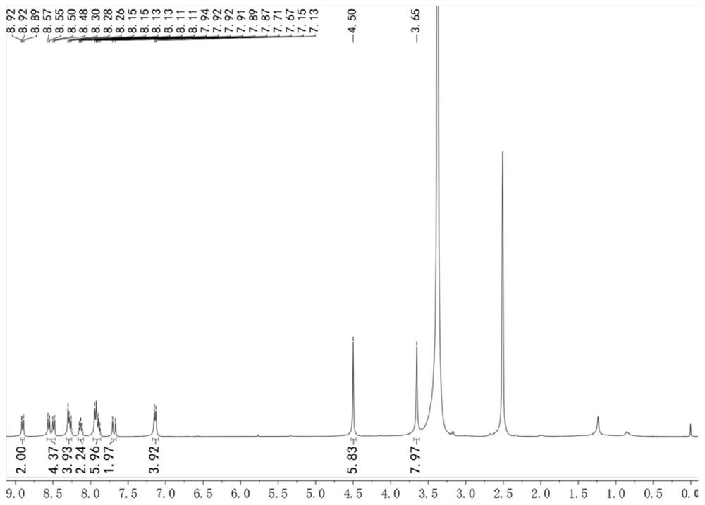
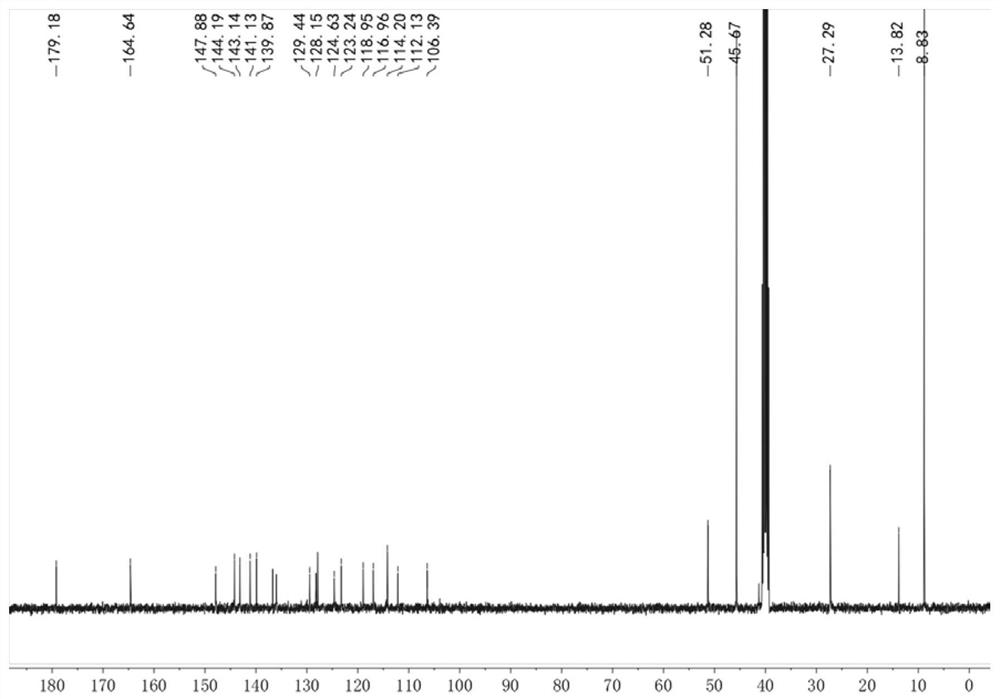
[0038] Add 10 mL of ethanol to the round-bottomed flask, then add 1.1 mL of 2-methylquinoline, add 0.5 mL of methyl iodide, and heat to 60 °C for 30 hours. After the reaction is completed, the reaction system is cooled to room temperature, and a solid is precipitated. , filtered and washed with ethanol to obtain 1,2-dimethyl-quinoline iodonium salt (compound 2) in 92% yield. 1 H NMR (400 MHz, DMSO-d6) δ 9.12 (d, J = 8.5 Hz, 1H), 8.60 (d, J = 9.0 Hz, 1H), 8.41 (dd, J = 8.2, 1.6 Hz, 1H), 8.24 (ddd, J = 8.8, 7.0, 1.6 Hz, 1H), 8.14 (d, J = 8.5 Hz, 1H), 8.00 (t, J = 7.6 Hz, 1H), 4.45 (s, 3H), 3.09 (s, 3H).

[0039] (2) Synthesis of 4,4'-(piperazine-1,4-diyl)benzaldehyde (compound 2):

[0040] Dissolve piperazine (1.0 g, 11.62 mmol) in H 2 O (18 mL) and 2-methoxyethanol (20 mL). The mixture was heated to reflux, then a solution of 4-fluorobenzaldehyde...

Embodiment 2

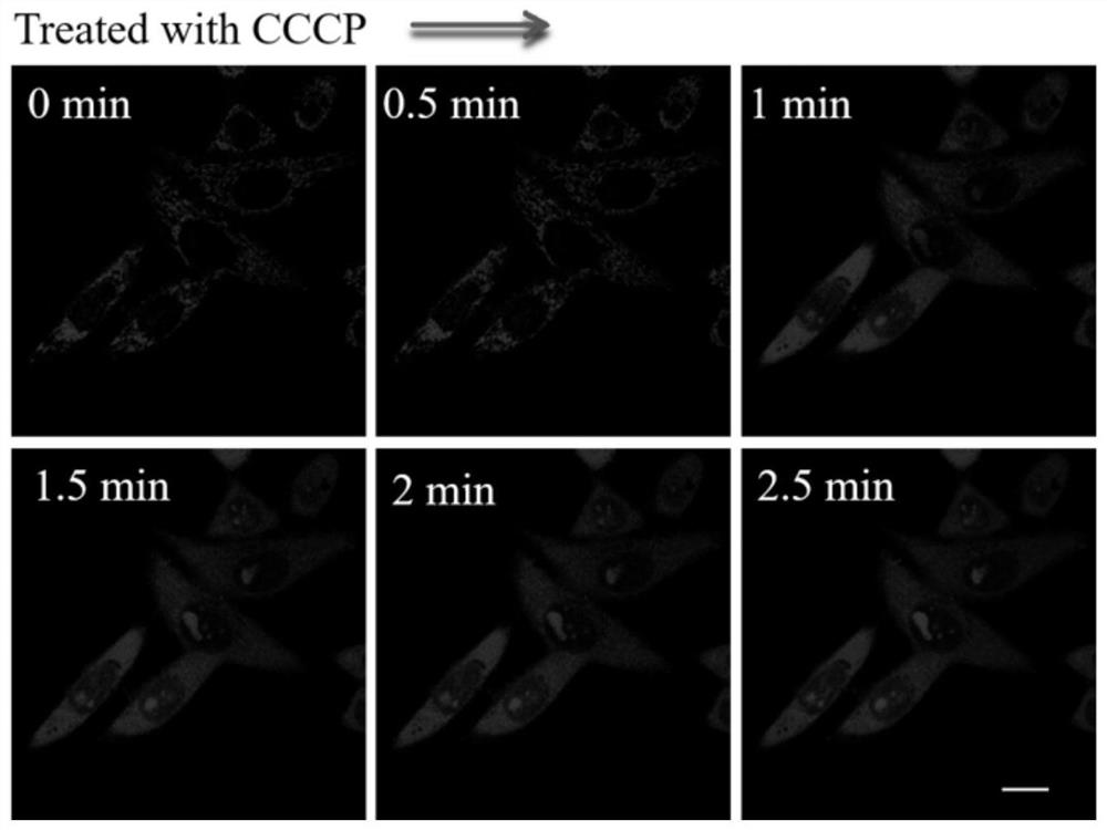
[0043] Example 2 Response of fluorescent probes to different membrane potentials

[0044] Set the density to 3 × 10 5 HeLa cells / mL were seeded into sterilized 35 mm imaging petri dishes under CO 2 Incubator (at 37 °C, 5 % CO 2 ) for more than 12 hours to make the cells adherent. Then, the DMSO solution of the probe HJI obtained in Example 1 with a concentration of 1 mM was prepared as the mother solution, and the mother solution was added to the cell culture dish to make the final concentration of 5 μM, and the culture was continued for 20 min, and then the cell culture solution was aspirated. Go, rinse the cells 3 times with PBS buffer, add 1 mL of fresh medium, and then add 10 μM CCCP (belongs to oxidative phosphorylation uncoupler, which can reduce mitochondrial membrane potential) Immediately image, every 0.5 minutes, record different Imaging pictures over time.

[0045] In the cell imaging experiment, the excitation wavelength was 488 nm, and the detection wavelengt...

Embodiment 3

[0046] Example 3 Selectivity of fluorescent probes for different ions

[0047] Prepare the DMSO stock solution of the fluorescent probe prepared in Example 1, the concentration is 5 mM, prepare different amino acids (Ile, Arg, Ser, Asn, Gln, Glu, His, Ala, Hcy, N-Ace, Val, GSH) and NaCl, KNO 3 , H 2 O 2 PBS stock solution at a concentration of 100 mM. Then add 5 μL of probe stock solution into 5 mL volumetric flasks, add 10 μL of different analytes to each volumetric flask, and finally make up to 5 mL with PBS buffer solution. Fluorescence detection was then performed (excitation wavelength 500 nm). Take the wavelength as the abscissa and the fluorescence intensity as the ordinate. Figure 4 . It can be seen from the figure that the addition of different analytes has no effect on the fluorescence of the probe.


PUM


Abstract

The invention belongs to the technical field of analytical chemistry and provides a fluorescent probe for detecting mitochondrial membrane potential, a preparation method and application thereof. The structural formula of the mitochondrial membrane potential fluorescent probe is: and can be obtained by reacting the reaction product of 4-fluorobenzaldehyde and piperazine with 1,2-dimethyl-quinoline iodide salt. The fluorescent probe provided by the invention has the characteristics of low biological toxicity, good membrane permeability, simple synthesis method, and simple purification steps, and the probe can be applied to cell imaging to detect, mark or display changes in mitochondrial membrane potential.

Description

technical field [0001] The invention belongs to the technical field of analytical chemistry, in particular to a fluorescent probe for detecting mitochondrial membrane potential and its application. Background technique [0002] During the mitochondrial tricarboxylic acid cycle, the protons inside the mitochondria are actively transported to the outside of the mitochondria, thus forming a mitochondrial membrane potential of -160mV~ -180mV, which is negative inside and outside positive. The mitochondrial membrane potential provides energy for the process of aerobic respiration, catalyzing its breakdown of compounds with high stability. In addition, the mitochondrial membrane potential is closely related to the cell state, and the level of mitochondrial membrane potential can accurately reflect the health of the cell. Therefore, real-time observation of changes in mitochondrial membrane potential has important physiological, pathological and pharmacological significance. [0...

Claims


Application Information

Patent Timeline
Patent Type & Authority Patents(China)
IPC IPC(8): C07D215/12C09K11/06G01N21/64
CPCC07D215/12C09K11/06C09K2211/1029C09K2211/1044G01N21/6428G01N21/6486
Inventor 林伟英郭丁一田明刚孙洁
Owner UNIV OF JINAN
Why Eureka
Try Eureka