A method for constructing a three-dimensional heart model based on ultrasound imaging

A technology of ultrasound imaging and three-dimensional models, applied in 3D modeling, neural learning methods, biological neural network models, etc., can solve the problems of consuming a lot of manpower and time, not being able to meet non-invasiveness, promotion restrictions, etc., and achieve high accuracy, Accurate heart volume and systolic function, the effect of great application value

Active Publication Date: 2020-08-18
杭州蔡卓医疗设备有限公司
View PDF2 Cites 0 Cited by
  • Summary
  • Abstract
  • Description
  • Claims
  • Application Information

AI Technical Summary

Problems solved by technology

[0005] The invention patent with application number CN200610077079.3 discloses a software product for three-dimensional cardiac imaging using ultrasonic contour reconstruction, which mainly uses manual input to mark the contours of interest related to features of anatomical structures in one or more ultrasonic images, and based on The contour of interest and the measured position and orientation coordinates construct a 3D model of the anatomical structure, but this method requires manual contour extraction, which consumes a lot of manpower and time, and is limited in promotion
[0006] The invention patent with the application number CN201910177198.3 discloses a three-dimensional model construction method, device and system. By inserting the ablation catheter into the inner cavity of the heart and positioning the ablation catheter against the inner wall of the heart, the inner contour of the heart can be accurately obtained. In this way, a three-dimensional model of the heart is constructed, but this method is complicated to operate and requires the built-in intervention of the ablation catheter, which cannot meet the requirements of non-invasiveness

Method used


Image

  • A method for constructing a three-dimensional heart model based on ultrasound imaging
  • A method for constructing a three-dimensional heart model based on ultrasound imaging
  • A method for constructing a three-dimensional heart model based on ultrasound imaging

Examples


Embodiment 1

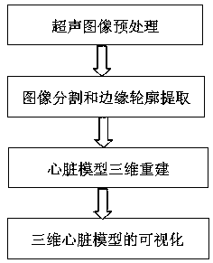
[0070] A method for constructing a three-dimensional heart model based on ultrasound imaging, comprising the steps of:

[0071] S1. Ultrasound image preprocessing: import the color Doppler cardiac ultrasound image data set, perform feature point recalibration processing on the data set, and then separate the data sets of each part of the heart (divided into left atrial data set, left ventricular data set, right Atrium dataset, right ventricle dataset, sinoatrial node dataset, atrioventricular node dataset, aortic dataset, pulmonary artery dataset, pulmonary vein dataset, superior and inferior vena cava dataset), ultrasonic speckle removal is performed for each dataset noise processing;

[0072] S2, image segmentation and edge contour feature extraction;

[0073] S3. Three-dimensional reconstruction of the heart model: the PTAM algorithm is used to carry out three-dimensional modeling of various parts of the heart, and then the generated single heart part models are synthesize...

Embodiment 2

[0100] A method for constructing a three-dimensional heart model based on ultrasound imaging, comprising the steps of:

[0101]S1. Ultrasound image preprocessing: import the color Doppler cardiac ultrasound image data set, perform feature point recalibration processing on the data set, and then separate the data sets of each part of the heart (divided into left atrial data set, left ventricular data set, right Atrium dataset, right ventricle dataset, sinoatrial node dataset, atrioventricular node dataset, aortic dataset, pulmonary artery dataset, pulmonary vein dataset, superior and inferior vena cava dataset), ultrasonic speckle removal is performed for each dataset noise processing;

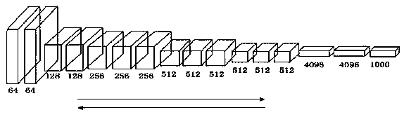
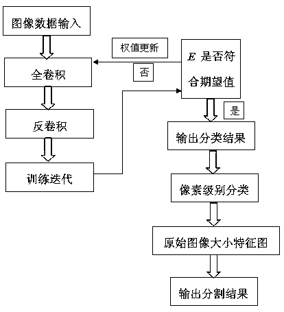
[0102] S2. Image segmentation and edge contour feature extraction: The improved full convolutional neural network learning algorithm is used to segment and edge contour extraction of cardiac ultrasound images, and the preprocessed cardiac ultrasound image datasets are respectively input to the ...


PUM


Abstract

The invention provides a method for constructing a three-dimensional heart model based on ultrasonic imaging. Firstly, the ultrasound images are preprocessed, and then the improved fully convolutional neural network learning algorithm is used to perform image segmentation and edge contour feature extraction on the data set, and the PTAM algorithm is used to detect and match the feature points of the cardiac ultrasound image and construct a three-dimensional heart model, and finally realize the visualization of the 3D heart model. The method provided by the invention has fine image segmentation effect, high feature extraction precision, fast model reconstruction speed, and high accuracy rate of the built three-dimensional heart model. The three-dimensional cardiac ultrasound model constructed by the present invention can be moved, rotated, and scaled arbitrarily in three-dimensional space, so as to observe the spatial position relationship of each tissue structure of the heart, thereby obtaining more diagnostic information than the two-dimensional cardiac ultrasound image, which has huge medical Value.

Description

technical field [0001] The invention relates to the technical field of three-dimensional modeling of ultrasonic images, in particular to a method for constructing a three-dimensional heart model based on ultrasonic imaging. Background technique [0002] Cardiovascular disease is the disease with the highest morbidity and mortality rate in the world, posing a serious threat to life and health. The early detection of the disease and the requirement of non-invasiveness in the diagnosis process have always been a difficult problem in the medical field. The heart is the main organ of the human circulatory system, and it is an extremely complex comprehensive system integrating electrophysiology, dynamics, and hemodynamics. [0003] The diagnosis and simulation of heart disease have been studied in depth. The early research mainly focused on the inverse problem of the heart, and analyzed and studied the cause of heart disease and the development of the disease through the existing...

Claims


Application Information

Patent Timeline
Patent Type & Authority Patents(China)
IPC IPC(8): G06T17/00G06T7/13G06T7/11G06T5/00G06N3/08G06N3/04
CPCG06T17/00G06T7/11G06T7/13G06T5/002G06N3/08G06T2207/10132G06T2207/30048G06N3/045
Inventor 张东海王宁
Owner 杭州蔡卓医疗设备有限公司
Why Eureka
Try Eureka