Super-resolution microscopy method and system based on saturated pumping-stimulated emission detection
A technology of stimulated radiation and microscopic system, which is applied in the super-resolution microscopic method and system field based on saturated pump-stimulated radiation detection, can solve the problem of signal strength weakening, achieve signal strength improvement, improve imaging speed, The effect of high imaging resolution
- Summary
- Abstract
- Description
- Claims
- Application Information
AI Technical Summary
Problems solved by technology
Method used
Image

Examples
Embodiment 1
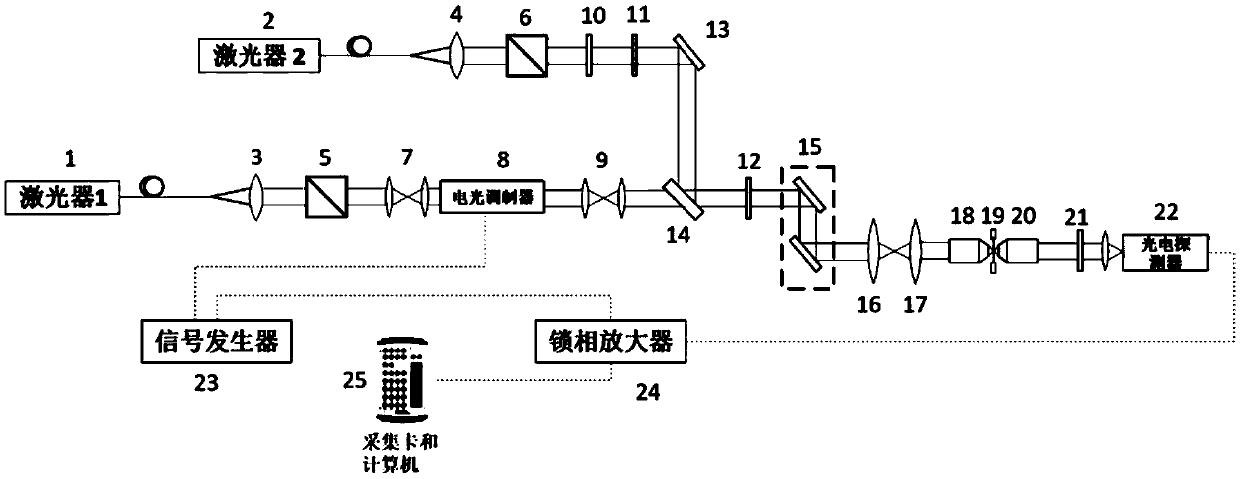
[0037] Such as figure 1 As shown, in this embodiment, a super-resolution microscopy system based on saturated pump-stimulated radiation detection for time-domain modulation by an electro-optical modulator includes a laser (wavelength λ 1 ) 1, laser 2 (wavelength λ 2 ), the first collimating lens 3, the second collimating lens 4, the first polarizing beam splitting prism 5, the second polarizing beam splitting prism 6, beam reducing mirror 7, electro-optic modulator 8, beam expanding mirror 9, 1 / 2 wave plate 10, 1 / 4 wave plate 11, 1 / 4 wave plate 12, mirror 13, dichroic mirror 14, scanning galvanometer 15, scanning mirror 16, field lens 17, illumination objective 18, sample 19, collection objective 20, filter Light sheet 21, photodetector 22, signal generator 23, lock-in amplifier 24, acquisition card and computer 25.
[0038] use figure 1 The system shown realizes super-resolution imaging based on saturated pump-stimulated radiation detection, and the process is as follows: ...
Embodiment 2
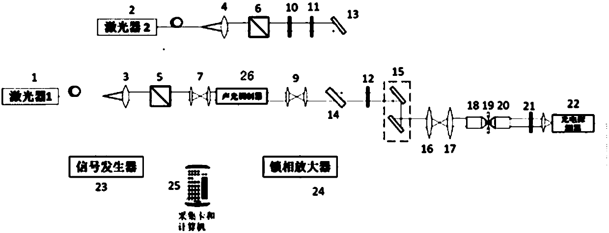
[0050] Such as image 3 As shown, in this embodiment, the super-resolution microscopy system based on saturated pump-stimulated radiation detection for time-domain modulation through an acousto-optic modulator, its structural composition is the same as that of the electro-optic modulator 28 except that the modulator is set as the Example 1 is the same. I won't repeat them here.
[0051] use image 3 The system shown realizes super-resolution imaging based on saturated pump-stimulated radiation detection, and the process is as follows:
[0052] (1) The wavelength emitted by laser 1 is λ 1 The excitation light is then collimated by the first collimating lens 3, and then converted into linearly polarized light by the first polarization beam splitter prism 5;
[0053] (2) The exciting light is narrowed by the beam shrinker 7, then the multi-level diffraction spot is obtained after the acousto-optic modulator 28, and the first-order diffracted light (time frequency is 10kHz), t...
Embodiment 3
[0063] This embodiment provides a super-resolution microscopy method based on saturated pump-stimulated radiation detection, including the following steps:
[0064] S1 The first light source emits a wavelength of λ 1 The excitation light is modulated in the time domain to make it loaded with a time frequency of 50kHz; the modulation device is an electro-optic modulator or an acousto-optic modulator.
[0065] S2 The second light source emits a wavelength of λ 2 the reading light.
[0066] S3 combining the excitation light with the time frequency and the reading light to form a combined beam.
[0067] S4 uses the combined beam to scan the sample, so that the sample emits stimulated radiation signal light.
[0068] The S5 stimulated emission signal light enters the photodetector after being collected and converted into an electrical signal. The method of backward detection is adopted, and the stimulated emission signal light is collected by the collection objective lens.
[...
PUM
 Abstract
Description
Claims
Application Information
 - Generate Ideas
- Intellectual Property
- Life Sciences
- Materials
- Tech Scout
- Unparalleled Data Quality
- Higher Quality Content
- 60% Fewer Hallucinations
Browse by: Latest US Patents, China's latest patents, Technical Efficacy Thesaurus, Application Domain, Technology Topic, Popular Technical Reports.
© 2025 PatSnap. All rights reserved.Legal|Privacy policy|Modern Slavery Act Transparency Statement|Sitemap|About US| Contact US: help@patsnap.com