Animal femoral surgery positioning and fixing device

A technique for surgical operation and fixation device, which is used in veterinary surgery, animal restraint instruments, medical science, etc., and can solve the problems of difficulty in realizing electric percutaneous needle insertion and bone piercing operation, limited operation space, and small femur size. , to achieve the effect of repeatable procedure standard and simplified surgical operation

Active Publication Date: 2020-03-20
HANGZHOU HUAMAI MEDICAL DEVICES CO LTD
View PDF6 Cites 2 Cited by
  • Summary
  • Abstract
  • Description
  • Claims
  • Application Information

AI Technical Summary

Problems solved by technology

Despite unremitting efforts, a novel ultra-thin cannula-type bone fixation needle was invented, which realized the possibility of electrically driving percutaneous bone fixation needles for the small bones of small animals. The operating space is limited, and the body of the anesthetized mouse is paralyzed, and the bone anatomy under the fur and soft tissue is difficult to distinguish, which still limits the possibility of bone fixation needle percutaneous puncture anchor needle operation
Practice has shown that it is very difficult to perform electric percutaneous needle insertion or accurate surgical operation without a standard three-dimensional surgical fixation system, and only rely on surgical assistants to stabilize the surgical bone with bare hands or hand-held bone forceps. needle piercing operation

Method used


Image

  • Animal femoral surgery positioning and fixing device
  • Animal femoral surgery positioning and fixing device
  • Animal femoral surgery positioning and fixing device

Examples


Embodiment 1

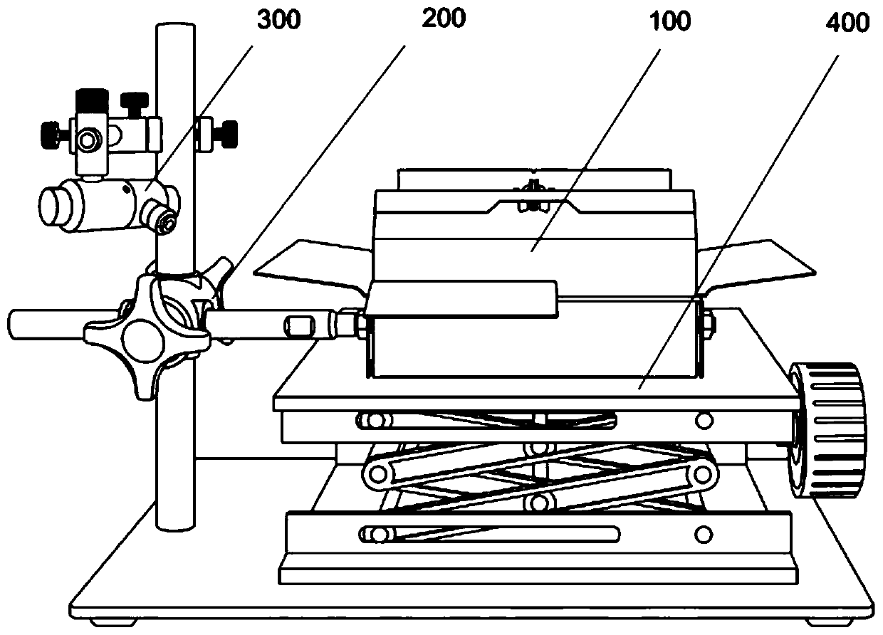
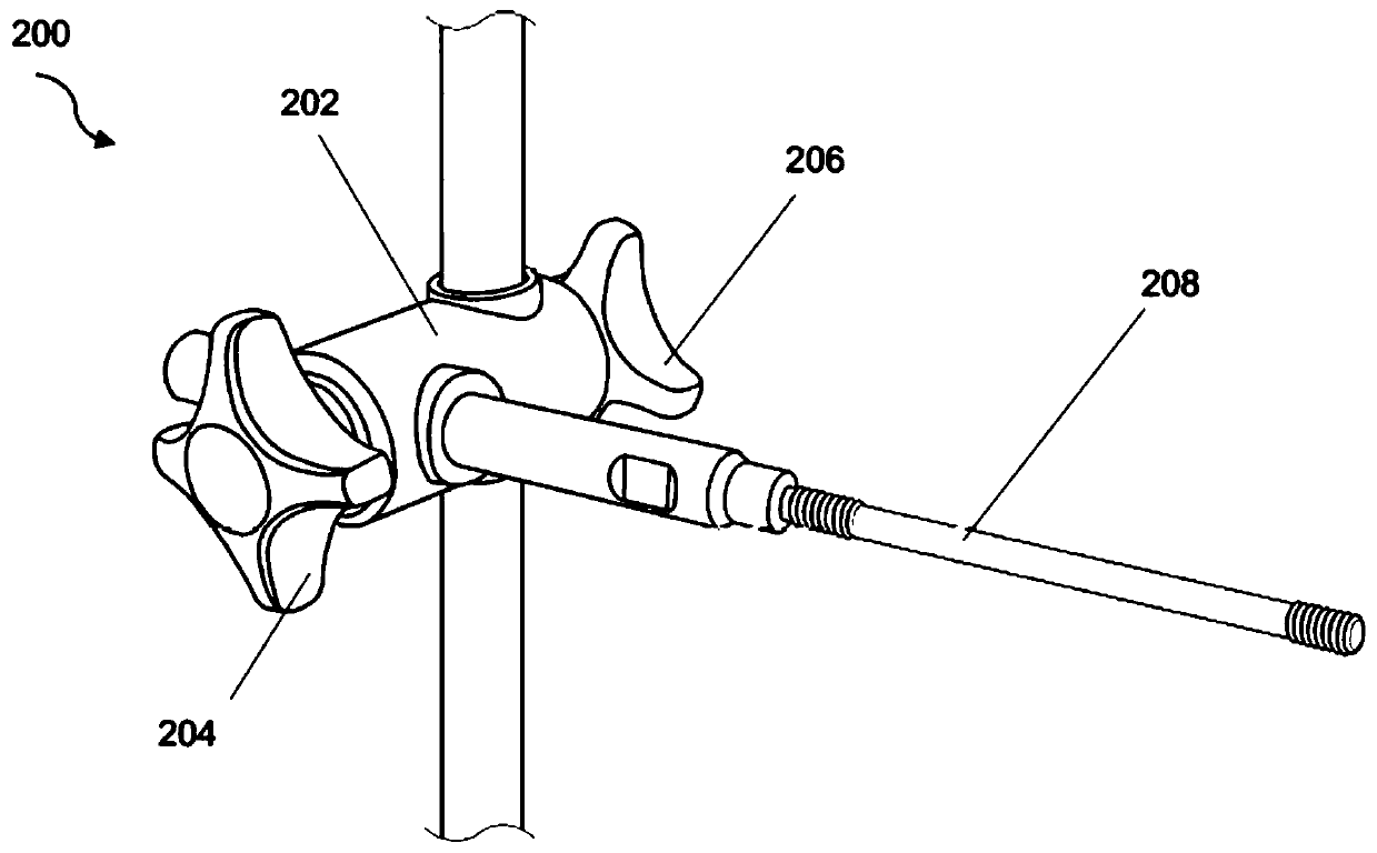
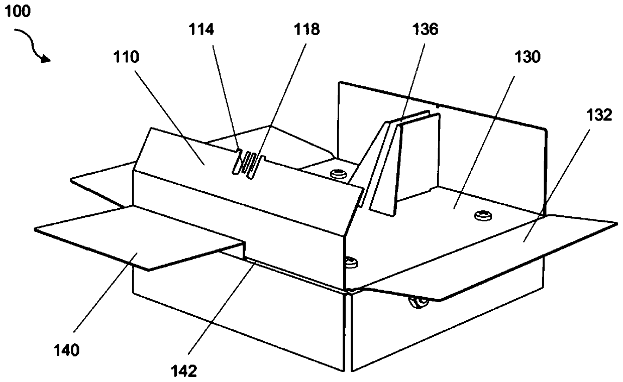
[0076] like Figure 1-15 The shown animal femur surgical positioning and fixing device includes a femoral positioning and fixing table 100. The femoral positioning and fixing table 100 is connected to an external three-dimensional fixation frame 200. The external three-dimensional fixation frame 200 is also connected with a laser marking and navigation unit 300. In addition, it is also equipped with The surgical lifting bracket 400 is used together with the femoral positioning and fixing table 100 . The external three-dimensional fixing frame is a three-dimensional three-dimensional adjustable fixing frame. The three-dimensional three-dimensional adjustable fixing frame is composed of a horizontal axis, a vertical axis and a three-dimensional three-dimensional regulator 202 connecting the horizontal axis and the vertical axis. The femoral positioning and fixing table 100 is composed of a fixing table deck 130 , a supine support plate 140 for an operating animal, a femoral pos...

Embodiment 2

[0105] like Figure 33 Shown is a schematic diagram of mouse femur fracture surgery using a rigid external fixator. Six needles penetrate the mouse femur and anchor the root in place; three needles at each end of the femur have outer segments bent parallel to each other toward the center to form shoulder bridges; fractures of the femur are generated with a bone breaker; coated with light-curable flowable composite The bridge is filled; the composite is cured with LED lights; the cured portion of the external fixator is removed after a few weeks by cutting the pins; all remaining pins are then spun out.

Embodiment 3

[0107] like Figure 34 Shown is a schematic diagram of mouse femur fracture surgery using a flexible external fixator. Six needles penetrated the mouse femur and anchored in place at the root; three distal femoral needles and three proximal femoral needles were each bent toward each other with three needles at each end in parallel to form two end cluster bridges; Each of them is coated with a light-curable flowable composite material and cured with an LED light; two elastic pins are placed and the two clusters at the two ends are connected step by step, that is, each left end and each right end of the two ends are connected and cured step by step through a light-curable flowable composite material. point; the right elastic pin is used to connect the right end faces of the two clusters, but only the proximal end is cured temporarily; the left elastic pin is used to connect the left end faces of the two clusters, but only the distal end is cured temporarily; the two elastic pins...


PUM


Abstract

The invention discloses an animal femoral surgery positioning and fixing device. The animal femoral surgery positioning and fixing device comprises a femoral positioning and fixing table, wherein thefemoral positioning and fixing table is provided with bone holding forceps for clamping an animal femoral shaft, a femoral positioning and fixing panel for fixing the neck of the bone holding forceps,and a fixing bin for fixing a handle of the bone holding forceps; and the femoral positioning and fixing table is used for limiting and fixing the surgical standard position of an animal femur in a femoral surgery, so that a surgical operation platform for a three-dimensional standard image of the femoral surgery is formed. By the animal femoral surgery positioning and fixing device, standardization of a femoral external fixation surgery for implementing experimental models such as femoral fracture, segmental bone defect and distraction osteogenesis can be guaranteed, so that a femoral modelsurgery technology has repeatability and data reliability. The animal femoral surgery positioning and fixing device is assisted by a three-dimensional digital fixing frame as an external fixing frame,and is linked with a synchronous laser marking navigation unit, so that a required large-environment suitable body position for the surgery is provided for a surgical operator, accurate positioning is realized, and meanwhile, a required basic environment is provided for a surgical robot operating system.

Description

technical field [0001] The invention relates to a special surgical instrument for medical experimental animal models, in particular to an animal femoral surgery technique invented for establishing models of femoral fractures, segmental bone defects and distraction osteogenesis in experimental animals and implementing orthopaedic external fixation techniques. The laser navigation positioning and fixation device can realize the standardization and repeatability of laboratory animal surgery, and provide a reliable guarantee for fixed-point digital surgery such as percutaneous puncture anchoring of bone fixation needles for external femoral fixation, femoral fractures or segmental bone defects. Background technique [0002] At present, the external fixation technology of small animal femoral experimental models in the international laboratory has always been a technical problem that many laboratories cannot hope to achieve. Due to the difficulty of implementing standard and repe...

Claims


Application Information

Patent Timeline
Patent Type & Authority Applications(China)
IPC IPC(8): A61D3/00A61D1/00
CPCA61D1/00A61D3/00A61D2003/006
Inventor 王力平江涛俞小华
Owner HANGZHOU HUAMAI MEDICAL DEVICES CO LTD
Why Eureka
Try Eureka