Application of TMED2 as treatment target spot of Ebola viral disease
An Ebola virus and target technology, applied in the field of biomedicine, can solve the problems of lack of vaccination options for treatment measures, increase of natural or man-made, infection of Ebola virus, etc.
- Summary
- Abstract
- Description
- Claims
- Application Information
AI Technical Summary
Problems solved by technology
Method used
Image
Examples
Embodiment 1
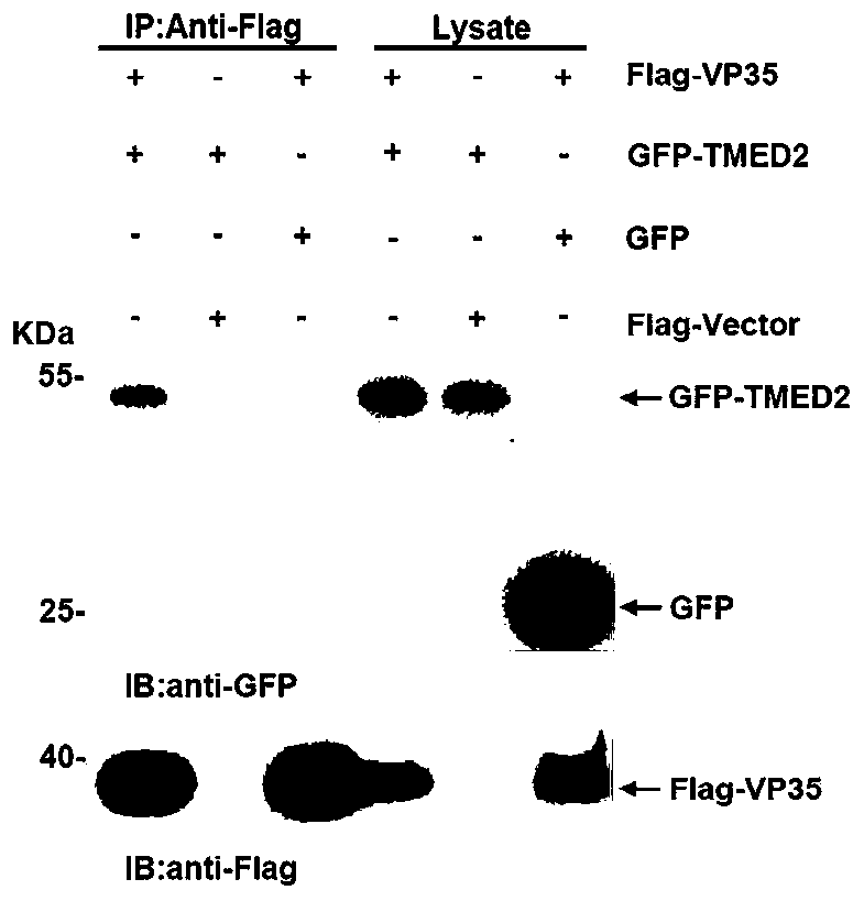
[0075] Example 1, immunoprecipitation and immunoblotting detection of the interaction between VP35 and TMED2
[0076] Co-transfect pcDNA3.0-Flag-VP35 and pEGFP-TMED2 plasmids in HEK293 cells, passage the cells into cell culture dishes as needed, and transfer the culture dishes into CO 2 Cultivate in an incubator, and transfect when the inoculated cells grow to 70-90% confluence, using Lipofectamine from Thermo Company TM 3000 transfection reagent, carry out plasmid transfection according to the manual: mix 1 μg pcDNA3.0-Flag-VP35 plasmid and 1 μg pEGFP-TMED2 plasmid with 4 μl P3000 at the ratio of plasmid:P3000=1:2 TM Add 100 μl Opti-MEM TM Dilute in culture medium to prepare DNA premix; mix 6 μl Lipofectamine according to plasmid: Lipo3000=1:3 TM Add 3000 to 100 μl Opti-MEM TM Diluted in the culture medium, in the diluted Lipofectamine TM Add the diluted DNA premix solution to the 3000 reagent, mix well and incubate at room temperature for 10-15 minutes, then add the DNA-...
Embodiment 2
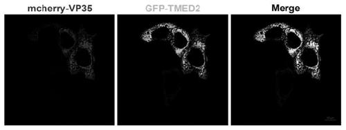
[0080] Example 2, live cell imaging detection of co-localization of VP35 and TMED2
[0081] Inoculate HepG cells into the chamber cover glass culture system one day in advance, and when the cells grow to a suitable density, transfect pcDNA3.0-mcherry-VP35 plasmid and pEGFP-TMED2 plasmid (for the operation method, see cell transfection), transfect After 24 hours of transfection, the chamber coverslip culture system was placed under a live cell imager to observe and take pictures.
[0082] The result is as figure 2 As shown, live-cell imaging technique found that VP35 co-localized with TMED2 in the cytoplasm.
Embodiment 3
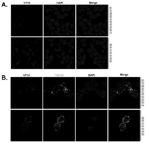
[0083] Example 3. Immunofluorescent staining detection of co-localization of VP35 and TMED2 in inclusion bodies
[0084] Inoculate HepG cells into a 6-well plate containing sterile coverslips one day in advance, and when the cells grow to a suitable density, transfect Ebola virus minimal genome plasmids: pCAGGS-NP (125ng), pCAGGS-VP35 (125ng) , pCAGGS-VP30 (75ng), pCAGGS-L (1000ng), p4cis-vRNA-Rluc (250ng) and pCAGGS-T7 (250ng) (see cell transfection for the operation method), after 48 hours of transfection, suck out the medium in the well , wash the cells 3 times with PBS at room temperature, and absorb the residual PBS for the last time; add 2ml 4% paraformaldehyde to each well and fix it at room temperature for 20-30min, absorb the paraformaldehyde, and wash the cells 3 times with PBS; add 2ml 0.3% Triton X-100 (prepared with 1×PBS) was perforated at room temperature for 15-20min, the Triton X-100 was aspirated, and the cells were washed 3 times with PBS; 2ml of blocking so...
PUM
 Abstract
Description
Claims
Application Information
 - R&D
- Intellectual Property
- Life Sciences
- Materials
- Tech Scout
- Unparalleled Data Quality
- Higher Quality Content
- 60% Fewer Hallucinations
Browse by: Latest US Patents, China's latest patents, Technical Efficacy Thesaurus, Application Domain, Technology Topic, Popular Technical Reports.
© 2025 PatSnap. All rights reserved.Legal|Privacy policy|Modern Slavery Act Transparency Statement|Sitemap|About US| Contact US: help@patsnap.com


