Biosensor for detecting exosome based on double aptamers, and production method and application thereof

A technology of biosensors and exosomes, applied in the field of biosensors, can solve the problems of cumbersome sample pretreatment process, and achieve the effects of low price, improved reaction speed, and low process cost

Active Publication Date: 2020-07-17
UNIV OF JINAN
View PDF6 Cites 5 Cited by
  • Summary
  • Abstract
  • Description
  • Claims
  • Application Information

AI Technical Summary

Problems solved by technology

[0004] In order to solve the problem of the cumbersome sample pretreatment process of exosome detection in the prior art, the present invention

Method used


Image

  • Biosensor for detecting exosome based on double aptamers, and production method and application thereof
  • Biosensor for detecting exosome based on double aptamers, and production method and application thereof
  • Biosensor for detecting exosome based on double aptamers, and production method and application thereof

Examples


Embodiment 1

[0050] The preparation method of described biosensor comprises the following steps:

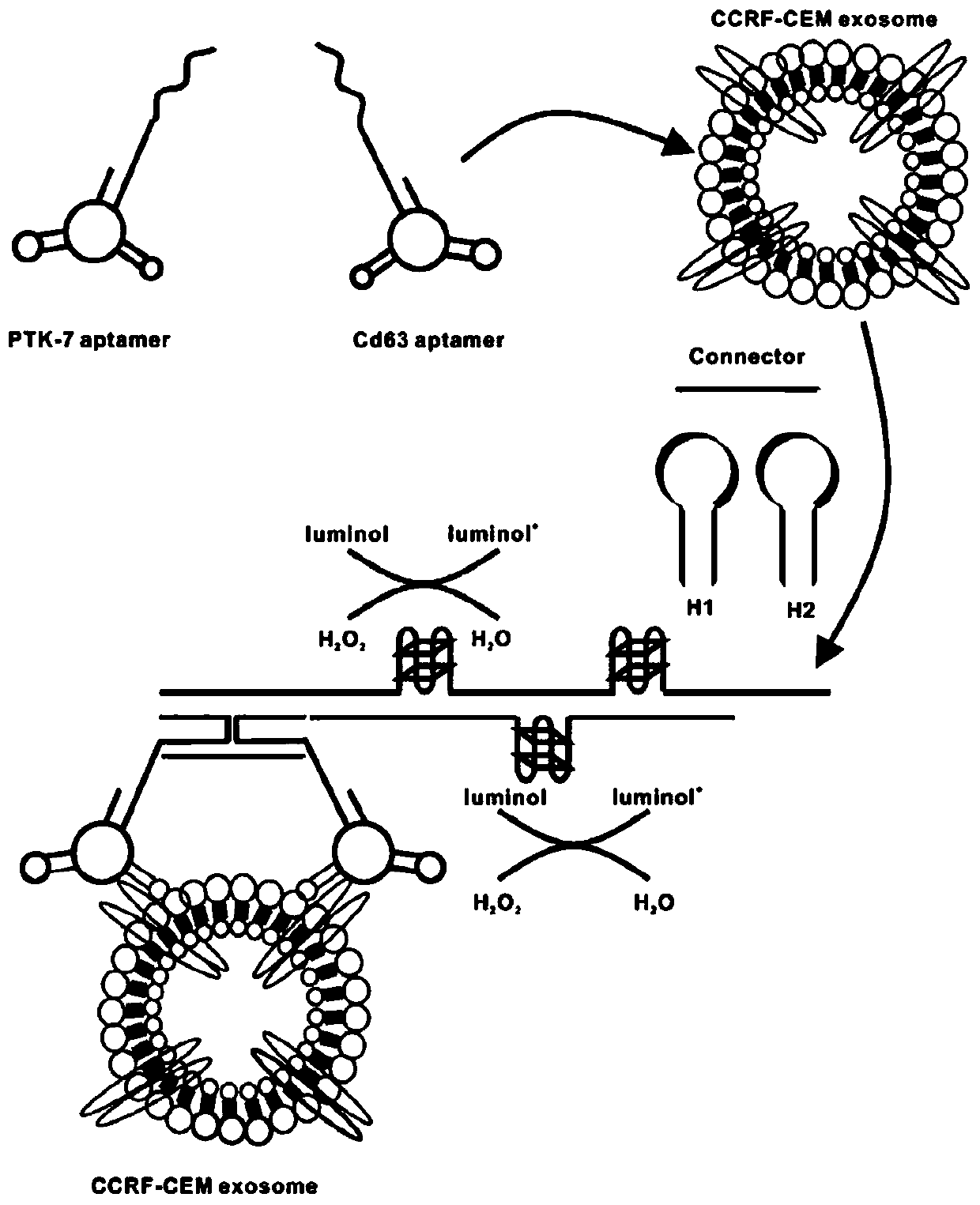
[0051] The operation steps of exosome extraction are as follows:

[0052] CCRF-CEM cell culture at 37 °C containing 5 % CO 2 in humid air. Passage every four days. RPMI-1640 4.5g / L, special grade fetal bovine serum (FBS), 1% antibiotics (100 U / mL penicillin and 100 μg / mL streptomycin), were used to culture the cells. To produce exosomes, cells were cultured in conditioned medium (10% FBS and 1% antibiotics) for three days and isolated using different ultracentrifugation methods. To isolate exosomes, cell debris was first removed from the culture medium by centrifugation at 2000 × g for 20 min. Cell vesicles were isolated by centrifugation at 10,000 × g for 45 min. Exosomes containing the supernatant were filtered with a 0.20-μm syringe filter. Finally, exosomes were harvested by centrifugation at 10,000 × g (40,000 rpm) for 150 min. The exosomes were redistributed in PBS and stored at ...

Embodiment 2

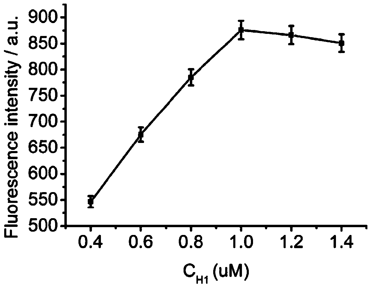
[0060] The preparation method of described biosensor comprises the following steps:

[0061] The operation steps of exosome extraction are as follows:

[0062] CCRF-CEM cell culture at 37 °C containing 5 % CO 2 in humid air. Passage every four days. RPMI-1640 4.5g / L, special grade fetal bovine serum (FBS), 1% antibiotics (100 U / mL penicillin and 100 μg / mL streptomycin), were used to culture the cells. To produce exosomes, cells were cultured in conditioned medium (10% FBS and 1% antibiotics) for three days and isolated using different ultracentrifugation methods. To isolate exosomes, cell debris was first removed from the culture medium by centrifugation at 2000 × g for 20 min. Cell vesicles were isolated by centrifugation at 10,000 × g for 45 min. Exosomes containing the supernatant were filtered with a 0.20-μm syringe filter. Finally, exosomes were harvested by centrifugation at 10,000 × g (40,000 rpm) for 150 min. The exosomes were redistributed in PBS and stored at ...

Embodiment 3

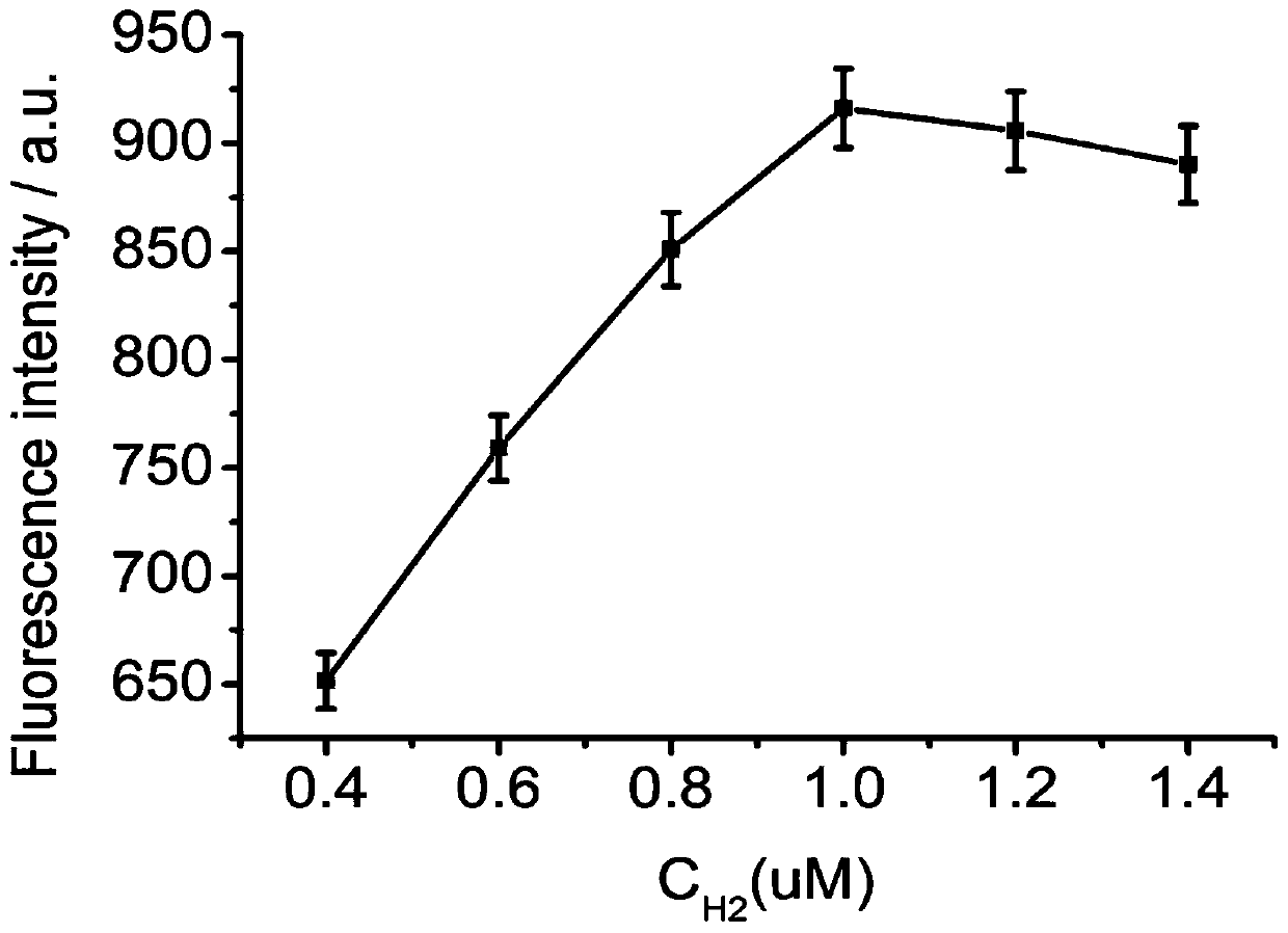
[0070] The preparation method of described biosensor comprises the following steps:

[0071] The operation steps of exosome extraction are as follows:

[0072] CCRF-CEM cell culture at 37 °C containing 5 % CO 2 in humid air. Passage every four days. RPMI-1640 4.5g / L, special grade fetal bovine serum (FBS), 1% antibiotics (100 U / mL penicillin and 100 μg / mL streptomycin), were used to culture the cells. To produce exosomes, cells were cultured in conditioned medium (10% FBS and 1% antibiotics) for three days and isolated using different ultracentrifugation methods. To isolate exosomes, cell debris was first removed from the culture medium by centrifugation at 2000 × g for 20 min. Cell vesicles were isolated by centrifugation at 10,000 × g for 45 min. Exosomes containing the supernatant were filtered with a 0.20-μm syringe filter. Finally, exosomes were harvested by centrifugation at 10,000 × g (40,000 rpm) for 150 min. The exosomes were redistributed in PBS and stored at ...


PUM

PropertyMeasurementUnit
Luminescence spectrumaaaaaaaaaa

Abstract

The invention relates to the technical field of biosensors, and particularly relates to a biosensor for detecting exosome based on a nucleic acid aptamer. The biosensor comprises an aptamer PTK-7 Apt,CD63 Apt, a connector, a hairpin probe H1, a hairpin probe H2 and a CCRF-CEM. The invention also relates to a production method and an application thereof. Based on specific recognition of the nucleic acid aptamer and a target object, through connection of the connector, two chains of PTK-7 Apt and CD63 Apt are adjacent to each other to form a trigger, and the trigger further opens H1 and the H1opens H2 to trigger an HCR reaction so that amplification of chemiluminescence intensity signals is realized, and the aptamer biosensor is constructed. The sensor has advantages of a high detection speed, simplicity in operation, a low price, a low detection limit, high specificity and the like.

Description

technical field [0001] The invention relates to the technical field of biosensors, in particular to a biosensor for detecting exosomes based on nucleic acid aptamers, and also relates to a preparation method thereof. Background technique [0002] Exosomes are membranous vesicles secreted by cells, usually with a diameter of 30–100 nm and a density of 1.10–1.18 Kg / L. Exosomes widely exist in various body fluids, can carry lipids, proteins, messenger RNAs, non-coding RNAs and other important biological functional molecules, and can participate in the exchange of substances and information between cells. Tumor-associated proteins, nucleic acids, and small molecules contained in exosomes have the potential to become potential biomarkers. The content of exosomes secreted by tumor cells is very high in the blood, with more than 10 exosomes per milliliter of blood 9 Exosomes, using exosomes as biomarkers for detection will have higher sensitivity, which is helpful for early detec...

Claims


Application Information

Patent Timeline
IPC IPC(8): G01N33/574G01N33/569G01N21/76C12Q1/682
CPCG01N33/57426G01N33/57492G01N33/56966C12Q1/682G01N21/76C12Q2525/301C12Q2525/205
Inventor 王玉孙文玉刘素黄加栋王业茹江龙张曼茹李莎莎王敬锋徐艺城
Owner UNIV OF JINAN
Why Eureka
Try Eureka