Respiration separation type strain imaging method based on ultrasonic images of living body

A technology of ultrasonic images and imaging methods, applied in the field of medical image processing, which can solve problems such as errors, increased errors in displacement and strain calculations, and gate failure

Active Publication Date: 2020-09-18
NANJING UNIV
View PDF5 Cites 2 Cited by
  • Summary
  • Abstract
  • Description
  • Claims
  • Application Information

AI Technical Summary

Problems solved by technology

However, the disadvantage of this program is that it can only choose one of the two states of "expiration" and "inhalation", that is, it can only output the results of one breathing sta

Method used


Image

  • Respiration separation type strain imaging method based on ultrasonic images of living body
  • Respiration separation type strain imaging method based on ultrasonic images of living body
  • Respiration separation type strain imaging method based on ultrasonic images of living body

Examples


Embodiment 1

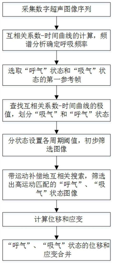
[0049] combine figure 1 Shown, a kind of respiratory separation type strain imaging method based on in vivo ultrasonic image of the present invention comprises the following steps:

[0050] 1) Acquisition of digital ultrasound image sequences

[0051] Ultrasonic image acquisition is performed on the strain-concentrated area of ​​the living body to obtain a digital ultrasonic image sequence; specifically, the target area of ​​the living body is heated or an external force is applied to form concentrated strain in the target area; it should be noted that the present invention is applicable to a variety of Application scenario requirements, such as tissue local temperature rise monitoring, elastic imaging, tissue scatterer motion tracking, etc. It is further worth noting that when an external action is applied to biological tissue, the specific form of the action is not limited, and it can be microwave ablation, radiofrequency ablation, ultrasonic ablation, laser ablation, and i...

Embodiment 2

[0091] In this example, the method of Example 1 is used to heat the adipose tissue of live pigs by microwave ablation, and the thermal strain is calculated. The specific steps of the method are as follows:

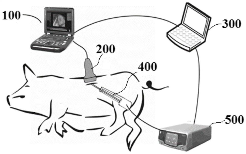
[0092] Step 1, such as figure 2 As shown, the ultrasonic imaging system 100 of the present embodiment adopts a B-ultrasound imager with a sampling rate of 40 MHz, and the imaging probe 200 is a linear array probe with 128 elements and a center frequency of 10.5 MHz to image live pig adipose tissue, with an imaging depth of 4 cm, microwave The power emission area of ​​the ablation needle 400 is on the imaging plane; the B-ultrasound imager outputs RF images with a size of 128 pixels*2048 pixels per frame at an average frame rate of 50 frames per second, and each experiment uses a microwave heater 500 from the microwave An image sequence with a length of 20 seconds was acquired from the start of heating. The computer 300 controls the ultrasonic instrument and the microwave...


PUM


Abstract

The invention discloses a respiration separation type strain imaging method based on ultrasonic images of a living body, and belongs to the field of medical image processing. The method comprises thesteps of acquiring a digital ultrasonic image sequence, obtaining the respiration and heartbeat frequency of a living body through two-dimensional cross-correlation calculation and spectrum analysis,then performing alternate extremum retrieval in a cross-correlation curve to divide the image sequence into an expiration state and an inspiration state, performing primary image screening, then respectively extracting an image sequence with the most matched motion state and a motion compensation amount corresponding to the image sequence, and calculating a spatial displacement and a spatial strain image sequence of a tissue according to the image sequence; and finally, combining the displacement image sequences and the strain image sequences of the two states respectively. The invention aimsto overcome the defects that physiological motions such as respiration and heartbeat of a living body influence speckle tracking precision to cause large artifacts and errors in a space strain image in the prior art, and the invention can obtain an accurate tissue internal displacement and strain distribution image.

Description

technical field [0001] The present invention relates to the field of medical image processing, and more specifically, relates to a breath-separated strain imaging method based on a living body ultrasonic image. Background technique [0002] With the development of medical imaging technology and the trend of minimally invasive and non-invasive treatment methods, medical imaging is more and more widely used in the monitoring of non-invasive and interventional treatment, typical examples include temperature imaging, elastic imaging and other functional imaging technologies in local tissue thermal ablation. . Commonly used imaging modalities include magnetic resonance imaging, computed tomography (CT) and ultrasound imaging. Ultrasound imaging equipment mainly based on B-ultrasound has been widely used in clinical practice due to its advantages of low cost, portability and strong real-time performance. [0003] When B-ultrasound is used for imaging in vivo, the ultrasonic imag...

Claims


Application Information

Patent Timeline
IPC IPC(8): G16H30/20G06T7/00
CPCG16H30/20G06T7/0012G06T2207/20104G06T2207/10132
Inventor 郭霞生尹楚豪章东屠娟
Owner NANJING UNIV
Why Eureka
Try Eureka