A kind of biological adhesive and its preparation method and application

A bio-adhesive, polyethylene glycol succinimide succinate technology, applied in the field of bio-adhesives and its preparation, can solve problems such as inability to suture effectively, poor repair effect, and inability to use alone. Achieve the effect of reducing surgical risk, shortening operating time, and good biocompatibility

Active Publication Date: 2022-07-29
BEIJING NATON INST OF MEDICAL TECH CO LTD
View PDF5 Cites 0 Cited by
  • Summary
  • Abstract
  • Description
  • Claims
  • Application Information

AI Technical Summary

Problems solved by technology

[0003] However, the existing artificial materials for dura mater repair generally have the problem of weak adhesion. They can only be used as auxiliary adhesives and cannot be used alone. They need to be covered on the surface of the dura mater after the dura mater is sutured to achieve watertight repair. Effect
In the actual application process, the location of the dura mater injury may be located in the front, side of the dura mater or the nerve root sleeve, etc. During the operation, it may not be possible to perform effective surgery due to the special injury site and poor dural suture technique. Even if artificial materials for dura mater repair are used, the repair effect is not good, and cerebrospinal fluid leakage still occurs from time to time after spinal surgery

Method used


Image

  • A kind of biological adhesive and its preparation method and application
  • A kind of biological adhesive and its preparation method and application

Examples


preparation example Construction

[0048] The preparation method of the bioadhesive of the present disclosure includes the following steps:

[0049] Step (1): The four-arm polyethylene glycol amino group is prepared into a first solution. The molecular weight of the four-arm polyethylene glycol amino group may be 2000-20000 Daltons.

[0050] In order to facilitate the use of the bioadhesive, the mass concentration of the four-arm polyethylene glycol amino group in the first solution in step (1) is 50 mg / mL-500 mg / mL.

[0051] Further, the mass concentration of the four-arm polyethylene glycol amino group in the first solution in step (1) is 100 mg / mL-300 mg / mL.

[0052] During specific implementation, the solvent of the first solution in step (1) may be one of secondary water, ultrapure water, physiological saline, or phosphate buffer with a pH of 7.4.

[0053] Step (2): Four-arm polyethylene glycol succinimidyl succinate is formulated into a second solution.

[0054] In order to facilitate the use of the bi...

Embodiment 1

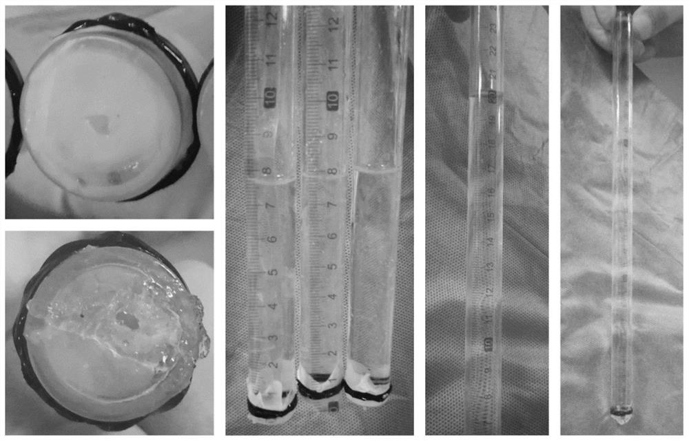
[0072] 400 mg of four-arm polyethylene glycol amino group (molecular weight of 20,000 Daltons) was weighed and dissolved in 2 mL of pure water to obtain the first solution. 300 mg of four-arm polyethylene glycol succinimidyl succinate (molecular weight: 20,000 Daltons) and 1.2 mg of genipin were weighed and dissolved in 2 mL of pure water to obtain a second solution. Use a double-barreled syringe to draw the first solution and the second solution, and inject the first solution and the second solution into the sample bottle at the same time, then invert the sample bottle, and record the gel time. The time when the gel does not flow back is the gelation time. The gel time was tested by the inversion method. The experimental results show that the gel formation time is 5-10 seconds, which meets the requirements of intraoperative operation.

Embodiment 2

[0074] 250 mg of four-arm polyethylene glycol amino group (molecular weight of 10,000 Daltons) was weighed and dissolved in 1 mL of pure water to obtain the first solution. 120 mg of four-arm polyethylene glycol succinimidyl succinate (molecular weight: 5000 Daltons) and 0.3 mg of genipin were weighed and dissolved in 1 mL of purified water to obtain a second solution. Use a double-barreled syringe to draw the first solution and the second solution, and inject the first solution and the second solution into the sample bottle at the same time, then invert the sample bottle, and record the gel time. The time when the gel does not flow back is the gelation time. The gel time was tested by the inversion method. The experimental results showed that the gel formation time was 5 to 20 seconds, which met the requirements of intraoperative operation.


PUM

PropertyMeasurementUnit
molecular weightaaaaaaaaaa
concentrationaaaaaaaaaa

Abstract

The invention discloses a biological adhesive and a preparation method and application thereof, wherein the biological adhesive comprises four-arm polyethylene glycol amino and four-arm polyethylene glycol succinate with a mass ratio of 1:(0.1-10). imidosuccinate. The bioadhesive of the present disclosure has good biocompatibility, is degradable, and has high adhesion ability, and is sufficient to withstand the pressure of cerebrospinal fluid after being adhered to the dura mater. Moreover, the bioadhesive of the present disclosure is convenient to operate during surgery, can be used alone without suture, greatly shortens the operation time, and reduces the operation risk. The bioadhesive of the present disclosure can be used for dural injuries in special parts that cannot be effectively sutured by conventional methods, such as injuries located in the anterior, lateral and nerve root sleeves of the dura mater, and can effectively reduce the complications of cerebrospinal fluid leakage after spinal surgery disease and improve patient outcomes.

Description

technical field [0001] The invention relates to the field of medical adhesives, and more particularly, to a biological adhesive and its preparation method and application. Background technique [0002] The current intraoperative repair methods for the dura mater mainly include direct suture method and dura mater replacement material suture method. Among them, the direct suture method can achieve the purpose of directly and effectively repairing the dura mater, but for those with dura mater defects during the operation, the direct suture often has too much tension, which is easy to form compression on the spinal cord, and because the suture needle during the suturing process is often too large. The resulting "eye of the needle" makes it difficult to achieve an absolute watertight suture. Using autologous or allogeneic tissue (such as muscle, fat, fascia or dura mater, etc.) as a dura mater substitute material and tension reduction suture can repair the dura mater with certai...

Claims


Application Information

Patent Timeline
Patent Type & Authority Patents(China)
IPC IPC(8): A61L31/06A61L31/14C08G81/00
CPCA61L31/041A61L31/14C08G81/00C08G2230/00C08L87/005
Inventor 祝腾蛟王星田耘
Owner BEIJING NATON INST OF MEDICAL TECH CO LTD
Why Eureka
Try Eureka