Device and method for screening and separating circulating tumor cells and application

A tumor cell and separation device technology, applied in the field of circulating tumor cell screening and separation devices, can solve the problems of low sensitivity and specificity, long time consumption, missed detection of CTCs, etc., to avoid loss of CTCs, avoid false negatives, and avoid false positives Effect

Active Publication Date: 2020-10-02
CIXI INST OF BIOMEDICAL ENG NINGBO INST OF MATERIALS TECH & ENG CHINESE ACAD OF SCI +1
View PDF7 Cites 2 Cited by
  • Summary
  • Abstract
  • Description
  • Claims
  • Application Information

AI Technical Summary

Problems solved by technology

However, this method requires a large amount of blood to screen and isolate CTCs, takes a long time, and has low sensitivity and specificity. During the operation, CTCs

Method used


Image

  • Device and method for screening and separating circulating tumor cells and application

Examples


Embodiment 1

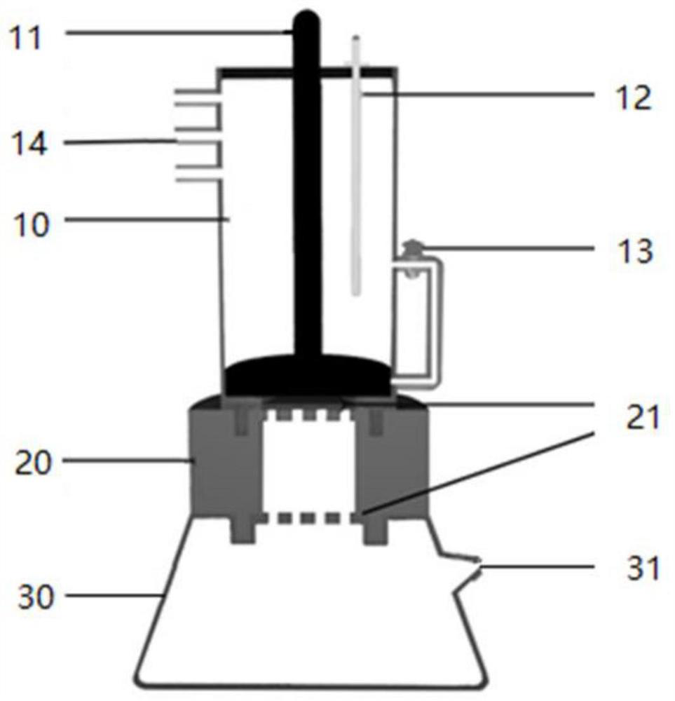
[0080] Select a blood sample incubation chamber with a height of 120 mm, an inner diameter of 15 mm, and an outer diameter of 16 mm, and a biofiltration membrane with a pore size of 0.5 to 1 micron, and connect the blood sample incubation chamber, cell screening chamber, and waste liquid recovery chamber. Inject nanomaterials and peripheral blood samples into chamber A of the blood sample incubation chamber, and after incubation for 1 hour, lift the piston push rod up to make the upper inlet of the catheter tube, so that the mixed solution enters chamber A. After the magnetite was used to attract and separate CTCs from the blood cells outside the blood sample incubation room, the mixed solution was sonicated for 30 seconds. Remove the isolation baffle between the blood sample incubation chamber and the cell screening chamber, and open the exhaust port of the waste liquid recovery chamber. Push the piston down at a constant speed until all the liquid enters the waste recovery c...

Embodiment 2

[0082] Select a blood sample incubation chamber with a height of 110 mm, an inner diameter of 18 mm, and an outer diameter of 21 mm, and a biofiltration membrane with a pore size of 0.5 to 1 micron, and connect the blood sample incubation chamber, the cell screening chamber and the waste liquid recovery chamber. Inject nanomaterials and peripheral blood samples into chamber A of the blood sample incubation chamber, and after incubation for 1 hour, lift the piston push rod up to make the upper inlet of the catheter tube, so that the mixed solution enters chamber A. After the magnetite was used to attract and separate CTCs from the blood cells outside the blood sample incubation room, the mixed solution was sonicated for 30 seconds. Remove the isolation baffle between the blood sample incubation chamber and the cell screening chamber, and open the exhaust port of the waste liquid recovery chamber. Push the piston down at a constant speed until all the liquid enters the waste rec...

Embodiment 3

[0084] Select a blood sample incubation chamber with a height of 100 mm, an inner diameter of 18 mm, and an outer diameter of 21 mm, and a biofiltration membrane with a pore size of 0.5 to 1 micron, and connect the blood sample incubation chamber, the cell screening chamber and the waste liquid recovery chamber. Inject nanomaterials and peripheral blood samples into chamber A of the blood sample incubation chamber, and after incubation for 1 hour, lift the piston push rod up to make the upper inlet of the catheter tube, so that the mixed solution enters chamber A. After the magnetite was used to attract and separate CTCs from the blood cells outside the blood sample incubation room, the mixed solution was sonicated for 30 seconds. Remove the isolation baffle between the blood sample incubation chamber and the cell screening chamber, and open the exhaust port of the waste liquid recovery chamber. Push the piston down at a constant speed until all the liquid enters the waste rec...


PUM

PropertyMeasurementUnit
Heightaaaaaaaaaa
The inside diameter ofaaaaaaaaaa
Outer diameteraaaaaaaaaa

Abstract

The invention discloses a device and method for screening and separating circulating tumor cells and application. The device comprises a blood sample culture chamber and a cell screening chamber whichare arranged from top to bottom; the blood sample culture chamber and the cell screening chamber are fixedly connected, and the cell screening chamber communicates with the blood sample culture chamber; the CTCs and blood cells in a blood sample are separated in the blood sample culture chamber by means of attraction action of a magnet; and biological filtering membranes are arranged in the cellscreening chamber, and the biological filtering membranes are used for intercepting the CTCs and filtering a nanometer material or the blood cells. By arranging the blood sample culture chamber, the CTCs and the blood cells can be separated in the blood sample culture chamber due to the fact that the magnetic nanometer material is attracted by the magnet, and CTCs loss caused by traditional CTCs separation by means of size difference is avoided, so that the detection accuracy is improved; and by arranging the cell screening chamber, the CTCs are filtered by the biological filtering membranes,so that fast screening and separation of the CTCs are achieved.

Description

technical field [0001] The application relates to a circulating tumor cell screening and separation device, method and application, belonging to the technical field of medical detection. Background technique [0002] The currently reported spectral detection technology is mainly based on surface-enhanced Raman scattering (SERS) detection of circulating tumor cells (Circulating Tumor Cells, CTCs), which is a rapid analysis of the presence of circulating tumor cells in peripheral blood. Methods, using highly specific and targeted SERS active nanomaterials with targeting ligands to complete the screening and identification of CTCs in blood cells. First, the peripheral blood sample was added to the peripheral blood lymphocyte separation medium, centrifuged at room temperature to stratify, and then the low-density cell layer containing leukocytes and CTCs was transferred to a new test tube, and the SERS active nanomaterials with the function of targeting CTCs were combined. Cult...

Claims


Application Information

Patent Timeline
IPC IPC(8): C12M1/42C12M1/12C12M1/00C12N5/09G01N21/65
CPCC12M47/04C12N5/0693G01N21/658C12N2509/00
Inventor 林杰吴爱国徐夏薇陈天翔何孟
Owner CIXI INST OF BIOMEDICAL ENG NINGBO INST OF MATERIALS TECH & ENG CHINESE ACAD OF SCI
Why Eureka
Try Eureka