Fluorescent protein and/or coupled protein monoclonal antibody labeling method and kit thereof

A monoclonal antibody and fluorescent protein technology, applied in the field of immunology, can solve the problems of poor signal-to-noise ratio of yin and yang signals and strong background signal, and achieve the effect of improving specificity

Active Publication Date: 2020-12-18
ZHEJIANG ZHENGXI BIOMEDICAL CO LTD
View PDF10 Cites 9 Cited by
  • Summary
  • Abstract
  • Description
  • Claims
  • Application Information

AI Technical Summary

Problems solved by technology

This method leads to poor signal-to-noise ratio and strong background signal of the final target protein PE marker when performing fluorescence detection.

Method used


Image

  • Fluorescent protein and/or coupled protein monoclonal antibody labeling method and kit thereof
  • Fluorescent protein and/or coupled protein monoclonal antibody labeling method and kit thereof
  • Fluorescent protein and/or coupled protein monoclonal antibody labeling method and kit thereof

Examples


Embodiment 1

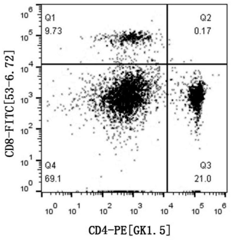
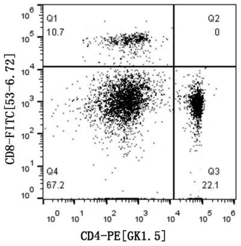
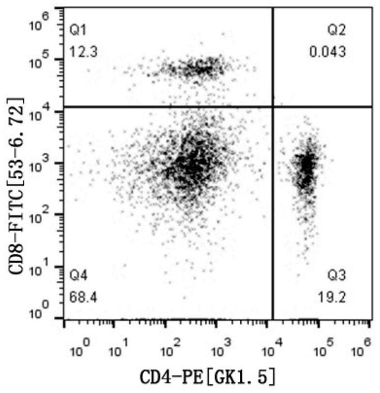
[0079] Example 1 Preparation of CD4[GK1.5]-PE (using the monoclonal antibody labeling method described in the present invention)

[0080] 1. Pretreatment of PE suspension

[0081] (1) Mix the PE suspension in the reagent tube with a vortex shaker.

[0082] (2) Take 0.5 mg of PE sediment suspension and place it in a 1.5 mL centrifuge tube, centrifuge at 12,000 g for 5 min, and completely absorb the supernatant with a pipette gun, taking care not to absorb the precipitate.

[0083] (3) Add 100 μL of PBS (containing 0.25 mM EDTA) buffer solution into the centrifuge tube to fully dissolve the PE precipitate at the bottom of the centrifuge tube, then centrifuge at 12000 g for 5 min, and absorb the PE supernatant solution.

[0084] (4) Select a 50Kd ultrafiltration centrifuge tube, move the PE supernatant solution into the ultrafiltration tube, then add 400 μL PBS (containing 0.25 mM EDTA) buffer and mix well, centrifuge at 12000 g for 5 min and remove the filtrate, then add 500 μL...

Embodiment 2

[0135] Example 2 Preparation of CD4[GK1.5]-PERCP (using the monoclonal antibody labeling method described in the present invention)

[0136] 1. PERCP sulfhydryl blocking reaction

[0137] (1) Take 50mM NEM out of the low temperature state and place it in room temperature environment. Open the bottle cap when the bottle temperature is balanced to room temperature to avoid condensation in the bottle.

[0138] (2) Add 7.5 μL 50 mM NEM solution (n PERCP :n NEM =1:50), reacted at 25°C for 1.5h, so that the NEM molecules combined with the free sulfhydryl groups in the PERCP molecules, and blocked the free sulfhydryl groups in the PERCP molecules.

[0139] 2. Cross-linking reaction between PERCP-NEM and S-SMCC

[0140] (1) Take S-SMCC (solubility 10mM, mother solution dissolved in sterile pure water) out of the low temperature state and place it in room temperature environment. Open the bottle cap when the temperature of the bottle is balanced to room temperature, so as not to app...


PUM


Abstract

The invention provides a fluorescent protein and / or coupled protein monoclonal antibody labeling method and a kit thereof. The preparation method comprises the following steps: firstly, carrying out desalination treatment on fluorescent protein and / or coupled protein, blocking sulfydryl of the fluorescent protein and / or coupled protein by adopting NEM, and carrying out cross-linking reaction on the fluorescent protein and / or coupled protein NEM and S-SMCC; meanwhile, cross-linking SLCSPDP and the monoclonal antibody, reducing disulfide bonds of the monoclonal antibody through TCEP, and conducting sulfhydrylation on the monoclonal antibody; and finally, carrying out cross-linking reaction on the sulfhydrylated antibody and NEM-fluorescent protein and / or coupled protein-S-SMCC. Directional coupling of the fluorescent protein and / or the coupling protein and the monoclonal antibody is achieved, the cross-linking efficiency of labeling of the fluorescent protein and / or the coupling proteinand the monoclonal antibody is improved, the specificity of labeling of the fluorescent protein and / or the coupling protein and the monoclonal antibody is further improved, and a fluorescence signal is enhanced.

Description

technical field [0001] The invention relates to the technical field of immunology, in particular to a fluorescent protein and / or coupling protein monoclonal antibody labeling method and a kit thereof. Background technique [0002] Monoclonal antibody is an antibody against a single antigenic determinant secreted by the fusion of mouse spleen immune cells and mouse myeloma cells to form hybridoma cells by using cell fusion technology and secreted under in vitro culture conditions. Under the stimulation of antigen, B lymphocytes can differentiate and proliferate to form specific antibodies against this antigen. The non-secretory myeloma cells grown in vitro can be fused with the B lymphocytes to form hybridoma cells, and then further cloned. The cloned hybridoma cells have the ability to produce specific antibody B lymphocytes It also has the ability of myeloma to grow infinitely in vitro, and a large amount of high-titer, single-specific antibody can be obtained by culturing...

Claims


Application Information

Patent Timeline
Patent Type & Authority Applications(China)
IPC IPC(8): G01N33/533G01N33/577G01N21/64
CPCG01N33/533G01N33/577G01N21/6486G01N21/6428G01N2333/70514G01N2333/70517G01N2333/7051G01N2021/6439
Inventor 张洋
Owner ZHEJIANG ZHENGXI BIOMEDICAL CO LTD
Why Eureka
Try Eureka