[0022]In order to make the above objectives, features, and advantages of the present invention more obvious and understandable, the specific embodiments of the present invention will be described in detail below in conjunction with specific embodiments.
[0023]In the following description, many specific details are explained in order to fully understand the present invention, but the present invention can also be implemented in other ways different from those described here, and those skilled in the art can do it without departing from the connotation of the present invention. Similar promotion, therefore, the present invention is not limited by the specific embodiments disclosed below.
[0024]Secondly, the “one embodiment” or “embodiment” referred to herein refers to a specific feature, structure, or characteristic that can be included in at least one implementation of the present invention. The appearances of "in one embodiment" in different places in this specification do not all refer to the same embodiment, nor are they separate or selectively mutually exclusive embodiments with other embodiments.
[0025]Experimental animals: 60 male SD rats, weighing about 140-160g, provided by the Animal Experiment Center of Xuzhou Medical University, clean grade, standard rearing. The experimental protocol was approved by the Animal Ethics Committee of Xuzhou Medical University.
[0026]Experimental drug grouping: A. Bacterial wall composite sponge: prepared with a mass concentration (g/ml) of 0.5% Staphylococcus aureus bacterial wall and 2.5% sodium alginate aqueous solution, totaling 24 mL, adding to a 48-well plate, 0.5 mL per well,- Pre-freeze at 20℃ and freeze-dry in a lyophilizer; the mass concentration (g/ml) is 1% gelatin, 1% chitosan, 0.5% calcium lactate mixed aqueous solution totaling 24mL, after mixing, add Staphylococcus aureus bacterial wall and seaweed In sodium sponge, 0.5mL per sponge, react for 30min, pre-freeze at -20°, and freeze-dry. B. Bacterial-free sponge: 2.5% sodium alginate solution, pre-freeze at -20°, freeze-dry in a freeze dryer; prepare a mixture of 1% gelatin, 1% chitosan, 0.5% calcium lactate, and add sodium alginate sponge In the middle, react for 30min, pre-freeze at -20°, and freeze-dry. C. Iodoform yarn.
[0027]The preparation method of 0.5% Staphylococcus aureus bacterial wall is: inoculate Staphylococcus aureus (ATCC6538) in liquid medium (inoculate 1×10 per liter)9cfu) Cultivate for 48h, divide the liquid medium suspension containing bacteria into centrifuge tubes, centrifuge at 12,000 rm for 20 minutes, discard the supernatant, take the precipitate, and use 10mMgSO4Wash with 10mM Tris-HCl solution of pH7.4, centrifuge at 12000rm for 20min, discard the supernatant, and add 10mMgSO4Pour the 10mM Tris-HCl solution of pH 7.4 into a beaker, sonicate for 20min, divide the suspension into a centrifuge tube, centrifuge for 5min at 2000rm, take the supernatant, centrifuge at 12000rm for 20min, take the precipitate, and place it in a centrifuge tube. Centrifuge at 12000rm for 20min and freeze-dry. 0.5 g of the freeze-dried product was re-dissolved in 100 mL of deionized water to obtain a 0.5% staphylococcus aureus bacterial wall solution.
[0028]Establishment of dry socket syndrome model in rats: SD rats were injected intraperitoneally with 4% chloral hydrate (1ml/100g). After anesthesia, the gingiva was separated with a probe, the upper and right first molars were protruded, and the alveolar fossa bone was destroyed by scraping. Wall, expand the wound. A cotton plug soaked with 1:1000 epinephrine hydrochloride is placed in the extraction socket for 1-2 minutes to constrict the blood vessels in the alveolar socket and stop bleeding, then put the cotton plug dipped in saturated Staphylococcus aureus strains Tooth extraction wound within 2min. After the animals regained consciousness, they returned to the animal room and kept them routinely after 0.5h without water. After 3-4 days of infection, the alveolar fossa of the tooth extraction wound of the rat was covered by a layer of yellow and low gray necrotic tissue, the surrounding mucosa was swollen, and the wound did not shrink significantly, which means the model was infected successfully.
[0029]Treatment: On the fourth day, the rats were randomly divided into 3 groups, each with 20 rats. Group A used a bacterial wall sponge, group B used a sponge without bacterial wall, and group C used iodoform gauze. One side of the rat was used as the blank group and not treated, and the other side was used as the treatment group. To remove spoilage and necrotic tissue, use cotton balls soaked in 3% hydrogen peroxide solution and physiological saline to clean the alveolar sockets until they are clean , And then put drugs in groups. Check the condition of the medicine in the wound after 3 days, and re-treat if it falls off. Take out the iodoform yarn at 1 week.
[0030]Observation indicators: 1. Tooth extraction wound area measurement: at 7d, 10d, and 14d after treatment, the wound healing was observed and recorded by naked eyes, the maximum length (L) and width (W) of the wound were measured, and the average of three measurements was taken, and the wound area changed S=L*W/(L0*W0)*100%. Observe the change of wound area to compare the recovery of soft tissue in each group.
[0031]2. He staining: fix with 4% paraformaldehyde for 2-3 days and decalcify with 10% EDTA decalcification solution for 2-3 weeks. During 2-3 days, the decalcification solution needs to be replaced. When the specimen is translucent to the naked eye and feels It is elastic, and the probe can smoothly penetrate into the bone tissue, which is the completion of decalcification. Embed in paraffin, make sagittal continuous slices along the long axis of the jaw, with a thickness of 5 μm. Each group of slices takes three basically the same positions for use. Observe the histological changes of the alveolar fossa under a light microscope.
[0032]3. Elisa determination: After 14 days of treatment of the dry socket model, homogenize the local tissue, centrifuge at 1000 rpm for 20 minutes, and take the supernatant. Take out the Elisa kit (enzyme-labeled biology), put it at room temperature and rewarm for 20 minutes, take 50μL of different concentrations of standards and add it to the well plate, and use the sample diluent to dilute the sample 4 times into the well plate, except for blank wells In addition, add 100μL of horseradish peroxidase (HRP)-labeled detection antibody to each of the standard wells and sample wells, seal the reaction wells with a sealing film, and incubate in a 37°C incubator for 60 minutes. After the incubation is complete, discard the liquid. Pat dry on absorbent paper, fill each hole with 20 times diluted washing solution, let stand for 1 min, shake off the washing solution, pat dry on absorbent paper, repeat the plate washing 5 times, wash the plate is completed, add substrate A, each hole B each 50μL, incubate at 37°C for 15min in the dark, and finally add 500μL of stop solution to each well, and measure the OD value of each well at 450nm within 15min.
[0033]4. PCR assay: inoculate mouse RAW264.7 macrophages in a 6-well plate, incubate the bacterial wall composite sponge, non-bacterial wall sponge and iodoform gauze strips with the macrophages for 24 hours, and add sponge or gauze to each well 0.1g. After 24h, add a proper amount of RNAisoplus and lyse the cells by pipetting, let it stand for 5 minutes, add 1/5 of the lysate volume of chloroform, shake and mix, then stand for 5 minutes, and centrifuge at 12000g for 15 minutes at 4°C. Take the supernatant, add 0.5-1 times the volume of the lysate with isopropanol, mix gently, let stand at room temperature for 10 minutes, centrifuge at 12000g for 10 minutes at 4°C, take the precipitate, and wash with the same volume of 75% ethanol as the lysate Precipitate, centrifuge at 7500g at 4°C for 5 minutes, discard the supernatant, take the pellet, and dry it at room temperature to obtain RNA extract. Dissolve the above RNA extract in a certain amount of ribozyme-free water, measure the RNA concentration in an RNA concentration analyzer, prepare a reverse transcription reaction solution according to the instructions of the reverse transcription kit, add a sample containing a certain amount of RNA, mix well, and reverse the RNA In the recording synthesizer, cDNA was synthesized at 37℃ for 15 minutes, 85℃, 5 seconds, 4℃ cycle once. According to a 10μL volume system, add cDNA samples, primers, fluorescent solution and ribozyme-free water to 96-well plates, seal the plate with sealing membrane, centrifuge at 3500 rpm, 4°C for 5 minutes, and detect the cytokine levels of each group using a PCR detector expression. The primer sequence is as follows:
[0034]
[0035]
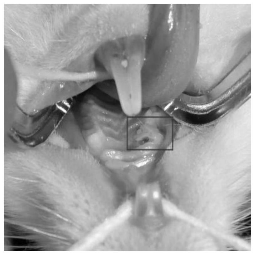
[0036]Visual observation: On the 4th day after tooth extraction, the surface of the alveolar socket was covered by a gray-yellow necrotic tissue layer, and the surrounding gums were red and swollen.figure 1 , Rat dry socket, the red area is the infection of the tooth extraction wound). The overall condition of the rat is slightly poor, the hair is less shiny than before, and the food intake is slightly reduced.
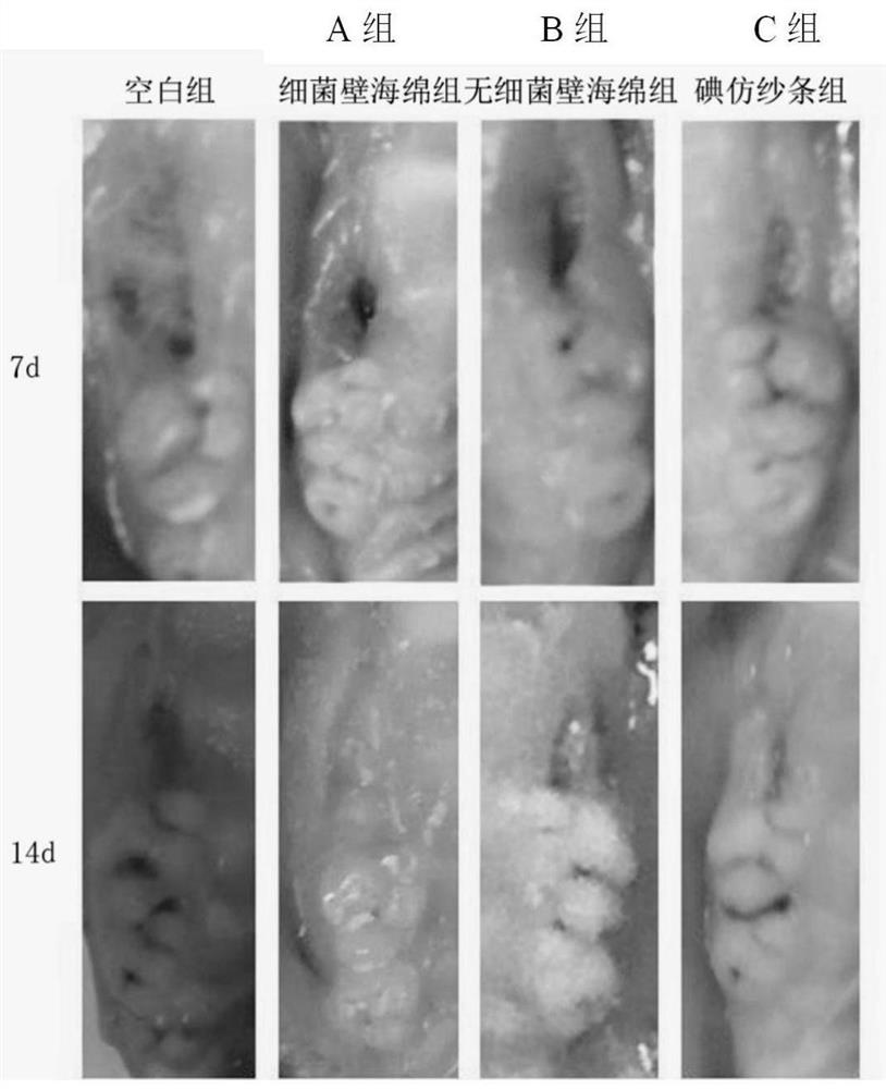
[0037]Observe after treatment,figure 2 It can be seen that the blank control group still has large wounds, and the wound inflammation in the treatment group decreased. The wounds healed gradually over time and the area decreased. At 7 days, the blank control group healed slowly, the area of the tooth extraction wound was still large, and the healing speed between the three treatment groups was bacterial wall sponge (group A)>No bacterial wall sponge group (group B)>Iodoform yarn group (C group). At 14 days, there were still obvious wounds in the blank control group, the wounds in the bacterial wall sponge group were basically completely healed, granulation tissue was visible in the tooth extraction wound, and there were still wounds in the non-bacterial wall sponge group and iodoform gauze group that did not heal.
[0038]Table 1 Measurement of tooth extraction wound area
[0039]
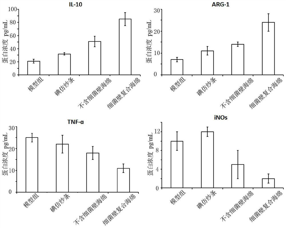
[0040]image 3 It is the result of Elisa examination of dry socket at 14 days. The results from the protein level of cytokines proved that compared with the iodoform gauze and the sponge without bacterial wall, the composite sponge with bacterial wall significantly increased the protein level of the anti-inflammatory factors (IL-10 and ARG-1) in the dry socket area and reduced The protein levels of its pro-inflammatory factors (TNF-ɑ and iNOs) indicate that the bacterial wall composite sponge has a good anti-inflammatory effect on dry socket disease.
[0041]Figure 4 In order to co-incubate the sponge with mouse RAW264.7 macrophages for 24h, the cell PCR inspection results. The results showed that compared with the iodoform gauze group (C group), the bacterial wall composite sponge group (A group) and the non-bacterial wall sponge group (B group) significantly increased the anti-inflammatory factors (IL-10 and IL-10) of RAW264.7 cells. The gene expression levels of ARG-1), the bacterial wall composite sponge group were significantly higher than the non-bacterial wall sponge group. The expression of pro-inflammatory factors (TNF-ɑ and iNOs) in the bacterial wall composite sponge group was significantly lower than the other three groups. The above results indicate that the bacterial wall composite sponge may change the type of macrophages, increase the RNA expression of macrophage anti-inflammatory factors, and reduce the RNA expression of pro-inflammatory factors, thereby exerting a therapeutic effect on dry socket disease.
[0042]Figure 5The results of HE staining of alveolar fossa tissue after the treatment of the bacterial wall sponge group and the blank control group. The blank control group had severe inflammation at 1 week. There were a large number of neutrophils in the alveolar fossa, lymphocyte infiltration and inflammatory infiltration on the surface. Out. The continuity of the epithelium in the composite sponge group was basically restored, the inflammation was significantly reduced compared with the blank control group, the scope was reduced, and fibrous connective tissue proliferation began to appear around it. At 2 weeks, the continuity of the mucosal epithelium of the blank control group was not fully recovered, while the composite sponge group had complete epithelium, more limited inflammation, and hyperplasia of fibrous connective tissue in the alveolar socket.
[0043]The present invention utilizes the typing changes of immune cells after bacterial infection and uses the bacterial wall of Staphylococcus aureus to prepare composite sponges. Research has found that the composite sponge prepared by the present invention can effectively regulate local macrophages and other immune cells, and promote tissue repair. The classification conversion can effectively avoid the inflammation caused by bacterial infection, thereby playing a good role in the treatment of dry socket disease and having good clinical application value.
[0044]It should be noted that the above embodiments are only used to illustrate the technical solutions of the present invention and not to limit them. Although the present invention has been described in detail with reference to the preferred embodiments, those of ordinary skill in the art should understand that the technical solutions of the present invention can be implemented Modifications or equivalent replacements without departing from the spirit and scope of the technical solution of the present invention should be covered by the scope of the claims of the present invention.