Umbilical cord blood Treg cell in-vitro amplification method based on trophoblastic cells and application

A trophoblast cell and cell body technology, applied in the field of biomedicine, can solve the problems of reduced cell activity, unfavorable clinical application, unfavorable Treg cell expansion in vitro, etc., and achieves the effect of reducing quality fluctuation, low immunogenicity and enhancing activity

Active Publication Date: 2021-03-09
成都云测医学生物技术有限公司
View PDF5 Cites 10 Cited by
  • Summary
  • Abstract
  • Description
  • Claims
  • Application Information

AI Technical Summary

Problems solved by technology

In the process of amplification, the expression of FoxP3 is also prone to instability, resulting in decreased cell viability, which is not conducive to clinical application
At the same time, due to the abnormal number and function of Treg cells in patients with autoimmune diseases, it is very unfavorable for the in vitro expansion of autologous Treg cells.

Method used


Image

  • Umbilical cord blood Treg cell in-vitro amplification method based on trophoblastic cells and application
  • Umbilical cord blood Treg cell in-vitro amplification method based on trophoblastic cells and application
  • Umbilical cord blood Treg cell in-vitro amplification method based on trophoblastic cells and application

Examples


Embodiment 1

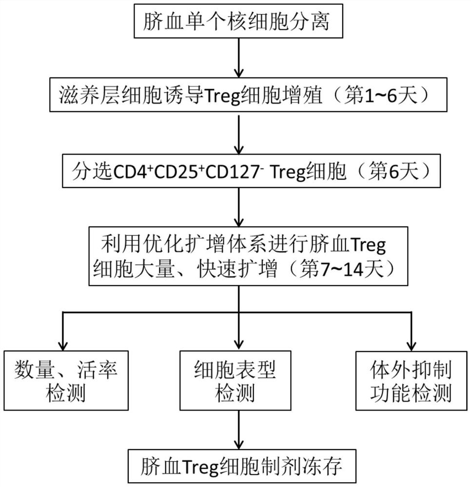
[0056] Prepare umbilical cord blood Treg cells according to the following steps, including the following steps:

[0057] Step 1: Collect 20 mL of fresh umbilical cord blood, dilute it with 2 times the volume of PBS, and then use density gradient centrifugation to separate umbilical cord blood mononuclear cells to obtain 6.2×10 7 cells.

[0058] Step 2: Adjust the mononuclear cells to 1 x 10 with culture medium 6 cells / mL, inoculate it into the culture flask; specifically, take 5×10 7 cord blood mononuclear cells, add 50mL X-VIVO 15 medium to resuspend and inoculate into T175 culture flask, and add 1×10 8 CD3 / CD28 immunomagnetic beads (the ratio of magnetic beads to cells is 2:1); add human AB plasma and IL-2 at the same time; after adding human AB plasma is 5vol%, and the final concentration of IL-2 is 500IU / mL.

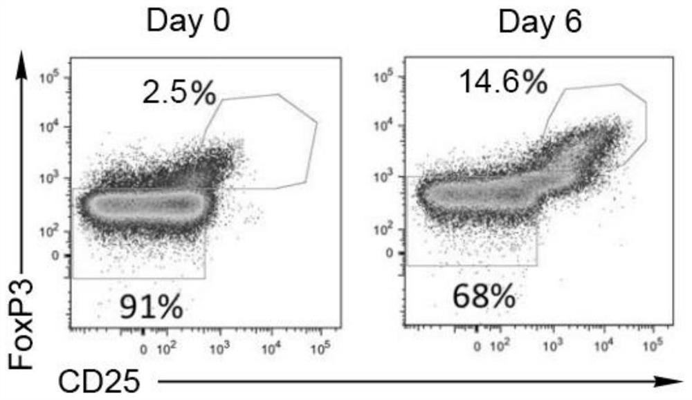
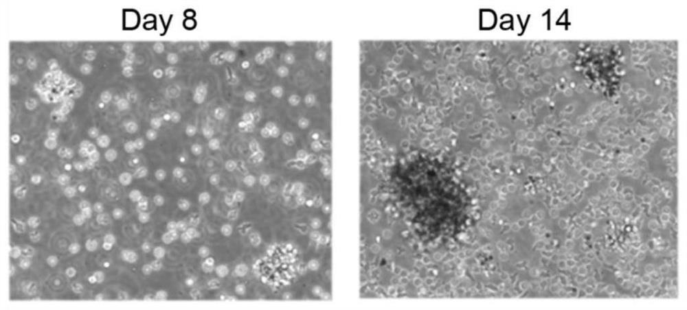
[0059] The 3rd generation umbilical cord Wharton's jelly mesenchymal stem cells were resuscitated as trophoblast cells and inoculated with 1×10 7 cells were co-c...

Embodiment 2

[0073] Cord blood Treg cells were prepared according to the same steps as in Example 1, except that the final concentration of each component after adding the optimized amplification factor in step 5 and step 6 was: human AB plasma was 10vol%, and the concentration of IL-2 was 1000IU / mL, the concentration of rapamycin is 100nmol / L, the concentration of RARA agonist is 10nmol / L, and the concentration of DNA methyltransferase inhibitor is 10μmol / L. The results show that the phenotype of Treg cells in this example is similar to that of Example 1, in which CD4 + CD25 + The positive rate was greater than 90%, and the positive rate of FoxP3 was greater than 80%. On the 14th day, the expansion factor of Treg cells was 3059.

Embodiment 3

[0075] Cord blood Treg cells were prepared according to the same steps as in Example 1, except that the final concentration of each component after adding the optimized amplification factor in step 5 and step 6 was: human AB plasma was 5vol%, and the concentration of IL-2 was 300IU / mL , the concentration of rapamycin was 100nmol / L, the concentration of RARA agonist was 1nmol / L, and the concentration of DNA methyltransferase inhibitor was 1μmol / L. The results show that the phenotype of Treg cells in this example is similar to that of Example 1, in which CD4 + CD25 + The positive rate was greater than 90%, and the positive rate of FoxP3 was greater than 80%. On the 14th day, the expansion factor of Treg cells was 1446.


PUM


Abstract

The invention discloses an umbilical cord blood Treg cell in-vitro amplification method based on trophoblastic cells and application. The specific technical method comprises the steps that firstly, umbilical cord Wharton's jelly mesenchymal stem cells are adopted as the trophoblastic cells to induce preliminary proliferation of Treg cells in umbilical cord blood mononuclear cells; then, pure Tregcells are obtained through magnetic bead sorting; and finally, the Treg cells are stimulated to be rapidly amplified by using optimized amplification factors. According to the amplification method, human AB plasma, IL-2, rapamycin, an RARA agonist and a DNA methyltransferase inhibitor are used as the optimized amplification factors, and a large number of umbilical cord blood Treg cells with high purity and high activity can be prepared within two weeks. Umbilical cord blood is used as a raw material for Treg cell amplification, batch preparation can be achieved, and Treg cell quality fluctuation caused by individual differences of samples can be reduced. The umbilical cord blood Treg cells have low immunogenicity and can be used as universal cells for clinical research, such as autoimmunediseases, graft-versus-host diseases and the like.

Description

technical field [0001] The invention relates to the technical field of biomedicine, in particular to a trophoblast cell-based umbilical cord blood Treg cell expansion method in vitro and its application. Background technique [0002] Regulatory T cells (Treg) are CD4 + An important cell subset in T cells, Treg cells play an important role in inducing the body's immune tolerance, maintaining the homeostasis of the immune environment, and preventing the occurrence of autoimmune diseases. Shimon Sakaguchi's team first identified CD25 (IL-2 receptor α-chain, IL-2RA) as a surface marker of Treg cells in 1995, and the researchers found that the removal of CD4 + CD25 + Transplantation of subsets of lymphocytes into nude mice can cause a variety of autoimmune diseases, while infusion of CD4 + CD25 + Treg cells can inhibit the occurrence of disease. At the same time, Treg cells also specifically express the transcription factor FoxP3, which plays an important regulatory role in ...

Claims


Application Information

Patent Timeline
Patent Type & Authority Applications(China)
IPC IPC(8): C12N5/0783A61K35/17A61P37/02A61P37/06A61P29/00A61P19/02A61P17/06A61P1/00
CPCA61K35/17A61P1/00A61P17/06A61P19/02A61P29/00A61P37/02A61P37/06C12N5/0637C12N2500/84C12N2501/04C12N2501/2302C12N2501/385C12N2501/51C12N2501/515C12N2501/72C12N2502/1388
Inventor 陈勇军刘少先
Owner 成都云测医学生物技术有限公司
Why Eureka
Try Eureka