Exosomes derived from granulocytic myeloid-derived suppressor cell and application thereof

A technology for inhibiting cells and granulocytes, applied in animal cells, tumor/cancer cells, vertebrate cells, etc., to achieve the effect of facilitating large-dose extraction, easy transportation, and promoting Treg polarization

Active Publication Date: 2015-06-03
JIANGSU UNIV
View PDF1 Cites 9 Cited by
  • Summary
  • Abstract
  • Description
  • Claims
  • Application Information

AI Technical Summary

Problems solved by technology

The application of G-MDSC exo in disease treatment has not been reported yet

Method used


Image

  • Exosomes derived from granulocytic myeloid-derived suppressor cell and application thereof
  • Exosomes derived from granulocytic myeloid-derived suppressor cell and application thereof
  • Exosomes derived from granulocytic myeloid-derived suppressor cell and application thereof

Examples


Embodiment 1

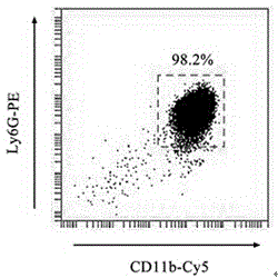
[0058] Example 1: Sorting of G-MDSCs and preparation of culture supernatant

[0059] (1) Establishment of Lewis lung cancer xenograft mouse model: Mouse Lewis lung adenocarcinoma cells preserved in our laboratory were cultured in DMEM medium containing 10% calf serum and pH 7.2 at 37°C and 5% CO2. With the cell growth density reaching 85% of the bottom area of ​​the culture dish as the standard, 0.25% trypsin was digested and passaged. With cells in logarithmic growth phase, 3.0×10 per mouse 6 The amount of cells was subcutaneously injected into 6-8w male C57BL / 6 mice in the right abdomen, and the growth of the tumor was observed after the tumor was planted.

[0060] (2) Establishment of the CIA model: Take an equal volume of bovine type II collagen (CII) and mix it with complete Freund's adjuvant at a ratio of 1:1, and grind it thoroughly until the mixture is completely emulsified. ). Take the emulsified CII and inject it intradermally at the base of the tail of ...

Embodiment 2

[0065] Example 2: Preparation of G-MDSC exo and determination of protein concentration

[0066] (1) Centrifuge the collected G-MDSCs supernatant at 4°C and 1000g for 30min to collect the supernatant; centrifuge the supernatant at 4°C and 10000g for 30min; transfer the supernatant to a MWCO 100 kDa ultrafiltration centrifuge tube and centrifuge at 1500g After 30 minutes, collect the concentrated solution in the inner tube.

[0067](2) Extract G-MDSC exo with the ExoQuick-TCTM Exosome kit purchased from SBI: the concentrated solution obtained in step (1) is mixed with the ExoQuick-TCTM Exosome reagent at a volume ratio of 5:1, shaken, and stand at 4°C More than 12 hours; centrifuge at 4°C, 1000g for 30 minutes, discard the supernatant, and collect the precipitate, which is G-MDSC exo. The prepared exosomes were dissolved in PBS, dispensed into EP tubes, and stored at -80°C for subsequent experiments.

[0068] (3) Determination of G-MDSC exo protein concentration with...

Embodiment 3

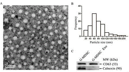
[0069] Example 3: Identification of G-MDSC exo

[0070] (1) Observation of G-MDSC exo morphology by transmission electron microscope: Take 20mL of G-MDSC exo suspension and drop it on a sample-loading copper grid with a diameter of 3 mm, and let it stand at room temperature for 2 minutes; gently blot the liquid with filter paper, and add 2 pH 6.8 dropwise. % phosphotungstic acid solution on the copper grid, negatively stained for 1min; filter paper to dry the dye solution, dried under an incandescent lamp, G-MDSC exo was observed under a transmission electron microscope as a round or elliptical microcapsule structure, with an envelope, cavity The inside is a low electron density component, the particle size is 30-150nm, the result is as follows figure 2 As shown in A and 2B, A is the morphology of G-MDSC exo. Under the transmission electron microscope, G-MDSC exo can be observed as a round or oval microcapsule structure with a complete envelope and low electron density ...


PUM

PropertyMeasurementUnit
particle diameteraaaaaaaaaa

Abstract

The invention relates to exosomes derived from granulocytic myeloid-derived suppressor cell and an application thereof, belongs to the fields of cytobiology, molecular biology and clinical application and particularly discloses a tumour individual or autoimmune disease individual (exosomes) (called G-MDSC exo) derived from the granulocytic myeloid-derived suppressor cell and application in preparation of medicine for inhibiting the autoimmune disease. The invention finds that the G-MDSC exo can effectively inhibit the CD4+T cell proliferation effect in vitro, promote the T regulartory cell induced proliferation and relieve the tardive hypersensitivity model mice foot swelling degree; the G-MDSC exo has inhibiting effects on the mice inflammatory bowel disease (IBD) and the collagen-induced arthritis (CIA). The application in preparation of medicine for effectively treating the autoimmune disease is also disclosed.

Description

technical field [0001] The invention relates to exosomes derived from granulocyte-like myeloid-derived suppressor cells and applications thereof, which belong to the field of cell biology, molecular biology and clinical application, and specifically relate to the preparation and biological Study on biological function and its application in the treatment of autoimmune diseases in mice. Background technique [0002] Myeloid-derived suppressor cells (MDSCs) are a group of heterogeneous cells derived from bone marrow, which are massively expanded in pathological conditions such as tumor, inflammation and pathogen infection (Gabrilovich DI, Nagaraj S. Myeloid-derived suppressor cells as regulators of the immune system. Nat Rev Immunol. 2009;9(3):162-74.). Mouse MDSCs are Gr-1 (composed of Ly6G and Ly6C) and CD11b double-positive cells, which can be divided into CD11b according to cell morphology and the expression of Ly6G and Ly6C + Ly6G + Ly6C low Granulocyte-like MDSCs (...

Claims


Application Information

Patent Timeline
Patent Type & Authority Applications(China)
IPC IPC(8): C12N5/078A61K35/15A61P37/02A61P29/00A61P19/02A61P37/08
CPCA61K35/15A61P19/02A61P29/00A61P37/02A61P37/08C12N5/0637C12N5/0693
Inventor 王胜军王运刚田洁芮棵马洁马斌汤新逸许化溪
Owner JIANGSU UNIV
Why Eureka
Try Eureka