Model for simulating dermatophyte infection in vitro and establishment method and application thereof

A dermatophyte, in vitro simulation technology, applied in the field of bioengineering, can solve the problems of dermatophyte reproduction, difficulty in triggering cell reactions, long and complicated construction process, etc., and achieve the effect of easy cellular immune response

Pending Publication Date: 2021-03-30
深圳钰捷生物医学科技有限公司
View PDF0 Cites 1 Cited by
  • Summary
  • Abstract
  • Description
  • Claims
  • Application Information

AI Technical Summary

Problems solved by technology

However, in the infection model established by using this structure, after dermatophyte infection, in the early stage of infection, due to the blocking of the outermost stratum corneum, it is difficult for the dermatophyte to directly contact the underlying cells, so it is difficult to trigger obvious cellular responses, which hinders Research on the cellular immune response to infection; and when the infection time is prolonged so that the fungus can penetrate the stratum corneum and directly contact the cells, it will cause a large number of fungus to multiply and adhere tightly to the stratum corneum of the epidermis Substances are not conducive to the use of high-throughput omics methods (such as high-throughput RNA seq

Method used


Image

  • Model for simulating dermatophyte infection in vitro and establishment method and application thereof
  • Model for simulating dermatophyte infection in vitro and establishment method and application thereof
  • Model for simulating dermatophyte infection in vitro and establishment method and application thereof

Examples


Embodiment 1

[0043] Example 1: Method of establishing an in vitro model simulating dermatophyte infection

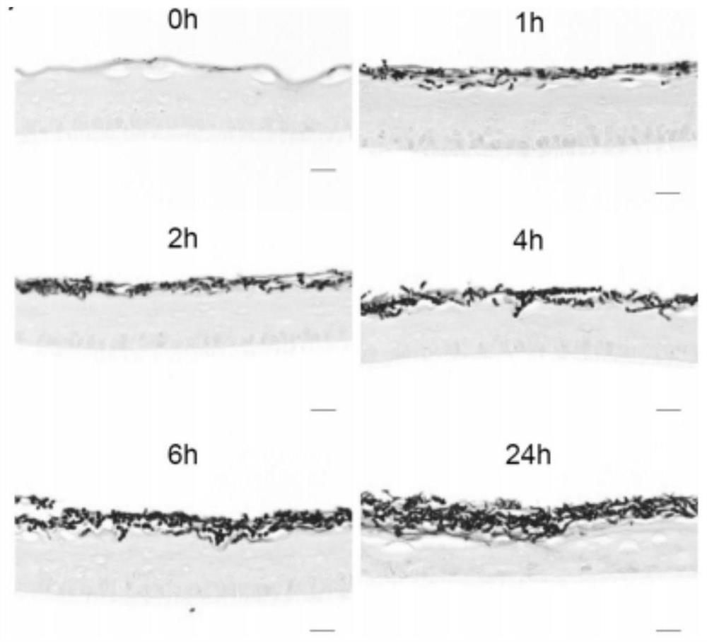
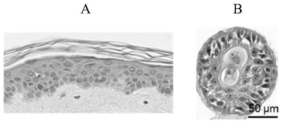
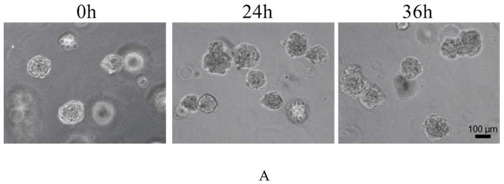
[0044] This embodiment establishes a kind of model that simulates dermatophyte infection in vitro, and the following description includes: the process of obtaining the cell spheres (also referred to as epidermis analogs herein) in the model, the process of obtaining dermatophyte conidia, and using the Infection of conidia with epidermal analogue cells to establish an in vitro model mimicking dermatophyte infection.

[0045] I. Acquisition of epidermis analogs

[0046] In this step, epidermal cells with differentiation function are cultured to obtain epidermal analogues. Epidermal stem / progenitor cells may be used from any commercial source or obtained using existing literature methods. The operation of culturing epidermal stem / progenitor cells isolated from human discarded foreskin tissue (the foreskin tissue comes from the Urology Department of the Fifth Medical Center of the Peop...

Embodiment 2

[0141] Example 2: Application of in vitro simulation of Trichophyton rubrum infection epidermis analog model in pathogenic mechanism research

[0142] This embodiment utilizes the in vitro simulated Trichophyton rubrum infection model established in the above-mentioned Example 1 (use the third medium to co-culture the Trichophyton rubrum conidia embedded in solidified matrigel and the epidermis analog for 24 hours to establish ), as the infection group; the epidermis analog embedded in the solidified Matrigel was simultaneously cultivated with the third medium for 24 hours, as the control group; the epidermis analog samples in the infection group and the control group were detected, and the red hair was analyzed. Pathogenesis of ringworm.

[0143] (1) Genomic sequencing of epidermal analogues in infected and control groups

[0144] High-throughput RNA sequencing (RNA-Seq) was performed on the epidermis analogs in the infection group and the control group, respectively, to stu...

Embodiment 3

[0183] Embodiment 3: the application of the model of simulating Trichophyton rubrum infection in vitro in the screening of anti-phytophyte drugs

[0184] This embodiment utilizes the in vitro simulated Trichophyton rubrum infection model established in the above-mentioned Example 1 (use the third medium to co-culture the Trichophyton rubrum conidia embedded in solidified matrigel and the epidermis analog for 24 hours to establish ) were tested for susceptibility to antiphytophyte drugs.

[0185] 1) The experimental group added amphotericin B (AmB) (a common anti-ringworm drug) to the model; the control group did not add amphotericin B to the model;

[0186] 2) After the experimental group and the control group were further cultured for 12 hours, the epidermal analogues of the experimental group and the control group were stained with glycogen granules (PAS) and observed under a transmission electron microscope according to operations 2.2 and 2.3 of Part III in the above-mentio...


PUM


Abstract

The invention discloses a model for simulating dermatophyte infection in vitro and an establishment method and application thereof, and belongs to the technical field of bioengineering. The model forsimulating dermatophyte infection in vitro comprises matrix glue, an epidermal analogue embedded in the solidified matrix glue and dermatophyte hyphae, the epidermal analogue is of a relatively complete solid sphere structure, and the exterior of the epidermal analogue is a cell layer which is composed of substrate layer cells and substrate upper layer cells and distributed in a surrounding mode,and a keratinization sample non-cell layer is arranged inside; hyphae invade basal layer cells outside the sphere structure and do not invade or only invade a small amount of basal layer cells, and donot invade the keratinization sample non-cell layer inside the sphere structure; the model can effectively simulate the state that dermatophyte infects normal skin tissue, the infection process of the model can be directly observed under a phase contrast microscope, the infection situation can be detected through omics methods such as high-throughput RNA sequencing, and the model can be effectively used for researching the pathogenic mechanism of dermatophyte infection and screening anti-dermatophyte drugs.

Description

technical field [0001] The invention belongs to the technical field of bioengineering, and specifically relates to an in vitro model for simulating dermatophyte infection and a method for establishing the same, as well as its application in the research of pathogenic mechanism of dermatophyte infection and in the screening of anti-dermatophyte drugs. Background technique [0002] Dermatophyte infection, commonly known as "beriberi", is the most common type of human fungal infection. The infection is usually limited to the epidermis, and most of it is caused by Trichophyton rubrum, which specifically eats human epidermal tissue. The clinical and pathological features of dermatophyte infection are chronic recurrent infection, thickening and exfoliation of keratinized skin tissue, and mild inflammatory response, but it is difficult for the human body to eradicate itself, and a good adaptability has been formed between the pathogen and the human host. Although not a serious life...

Claims


Application Information

Patent Timeline
IPC IPC(8): C12N5/071C12N1/14C12Q1/18C12R1/645
CPCC12N5/0625C12N1/14G01N33/5044C12N2500/32C12N2501/11C12N2501/70C12N2501/415C12N2503/06C12N2503/02C12N2533/90C12N2513/00G01N2500/10
Inventor 王韫芳王璇张伟宿钰鑫
Owner 深圳钰捷生物医学科技有限公司
Why Eureka
Try Eureka