Bionic nano-drug for preventing and treating aortic dissection and preparation method thereof

An aortic dissection, bionic nanotechnology, applied in the direction of drug combination, pharmaceutical formula, medical preparations with inactive ingredients, etc., can solve the problem of lack of effective therapeutic drugs, etc. High inflammatory targeting effect

Active Publication Date: 2021-04-30
FUDAN UNIV
View PDF0 Cites 2 Cited by
  • Summary
  • Abstract
  • Description
  • Claims
  • Application Information

AI Technical Summary

Problems solved by technology

Clinical practice shows that there is currently a lack of effective therapeutic drugs in clinical practice, and the search for preventive and therapeutic drugs and therapies to improve the level of prevention and treatment has attracted the attention of people in the industry

Method used


Image

  • Bionic nano-drug for preventing and treating aortic dissection and preparation method thereof
  • Bionic nano-drug for preventing and treating aortic dissection and preparation method thereof
  • Bionic nano-drug for preventing and treating aortic dissection and preparation method thereof

Examples


Embodiment 1

[0053] 1. Synthesis of DSPE-PEG2000-TN:

[0054] Weigh 2μM TN (YGRKKRRQRRRG-S-S-TTLDWSWLQMEC), dissolve in 1ml ddH 2 O, is solution A; weigh 1μM DSPE-PEG2000-MAL, dissolve in 2ml ddH 2 O, for solution B. Slowly add liquid B dropwise to liquid A under stirring, stir and react at room temperature for 12 hours, and freeze-dry the solution to obtain a white loose powder, which is DSPE-PEG2000-TN.

[0055] 2. Preparation of TN-(CUR)LP

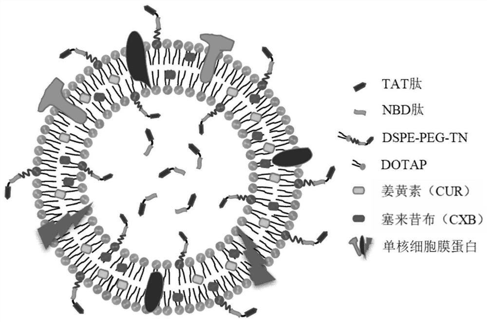
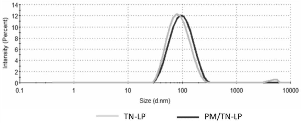
[0056] Weigh 24mg of SPC, 5mg of DOTAP, 3.6mg of cholesterol, 5.4mg of DSPE-PEG2000-TN, and 3mg of CUR into a round bottom flask, dissolve in 3ml of chloroform-methanol (4:1) mixed solvent, and remove the organic solvent by rotary evaporation at 40°C , forming a uniform lipid film on the inner wall of the flask. Add PBS 5.7 containing 0.3% Tween-80 to the lipid film, hydrate at 40°C for 30 minutes, and then sonicate at 80W for 3 minutes to obtain a liposome solution with a uniform particle size distribution of 80-100nm. Centrifuge at 10,000g for...

Embodiment 2

[0061] With the same preparation method as in Example 1, the CUR in the prescription was changed to the same dose of CUR and CXB, and the dosage of other components in the prescription was fixed to prepare PM / TN-(CUR&CXB)LP.

Embodiment 3

[0063] The preparation method is the same as in Example 1, and the PM in the prescription is changed to M1-type macrophage membrane protein purified by the same method. First, LPS is used to induce the polarization of monocytes into M1-type macrophages in vitro, and then the membrane protein is purified. , to fix the dosage of other components in the prescription, to obtain PM M1 / TN-(CUR / CXB)LP.


PUM

PropertyMeasurementUnit
particle sizeaaaaaaaaaa

Abstract

The invention belongs to the technical field of pharmaceutical preparations and nanomedicine, and relates to a bionic nano-drug for preventing and treating aortic dissection, in particular to a mononuclear/macrophage membrane bionic modified multidrug co-loaded anti-inflammatory liposome and a preparation method thereof. The liposome is composed of cationic phospholipid and neutral phospholipid, co-loading of multiple anti-inflammatory drugs can be achieved in the liposome, anti-inflammatory cell-penetrating peptide is covalently modified on the surface of the liposome, and then mononuclear/macrophage membrane protein is used for bionic modification. According to the bionic anti-inflammatory liposome, multiple receptors and adhesion molecular ligands mediated and chemotactic on the surface of a mononuclear/macrophage membrane are utilized to realize a natural targeting effect on an inflammatory site, and non-specific clearance of a reticuloendothelial system (RES) to the liposome is reduced by virtue of modification of leukocyte self-recognition molecules. The bionic anti-inflammatory lipidosome can effectively cross a vascular endothelial barrier, gather and release anti-inflammatory drugs at the early lesion part of the aortic dissection in a targeted manner, play a synergistic anti-inflammatory role, inhibit inflammation-related cells from chemotaxis and infiltration into the lesion part, and further prevent and treat the occurrence and development of the aortic dissection.

Description

technical field [0001] The invention belongs to the technical field of pharmaceutical preparations and nanomedicine, and relates to a biomimetic nano-medicine for preventing and treating aortic dissection, in particular to a mononuclear / macrophage membrane biomimetic modified multi-drug co-loaded anti-inflammatory liposome and a preparation method thereof. Background technique [0002] The prior art discloses that aortic dissection (Aortic Dissection) is a very dangerous and high-mortality macrovascular disease clinically. Studies have shown that in the course of the disease, vascular endothelial lesions caused by various reasons first cause aortic dissection. The intima of the wall forms a tear, and the blood in the aortic lumen, driven by the arterial pressure, enters the media through the tear, separates the media and expands along the long axis of the aorta, forming a false channel. Clinical practice shows that there is currently a lack of effective therapeutic drugs in ...

Claims


Application Information

Patent Timeline
Patent Type & Authority Applications(China)
IPC IPC(8): A61K9/127A61K47/24A61K47/28A61K47/46A61K38/08A61K38/10A61P29/00A61P9/00A61K31/12A61K31/635
CPCA61K9/127A61K47/24A61K47/28A61K47/46A61K38/08A61K38/10A61K31/12A61K31/635A61P29/00A61P9/00A61K2300/00
Inventor 姜嫣嫣刘敬璇吴俊龙刘晓
Owner FUDAN UNIV
Why Eureka
Try Eureka