

Precise immunofluorescence labeling method for integral tissue sample
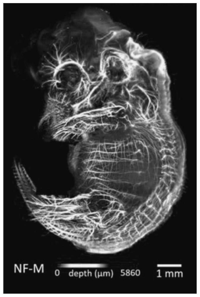
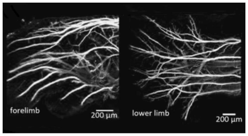
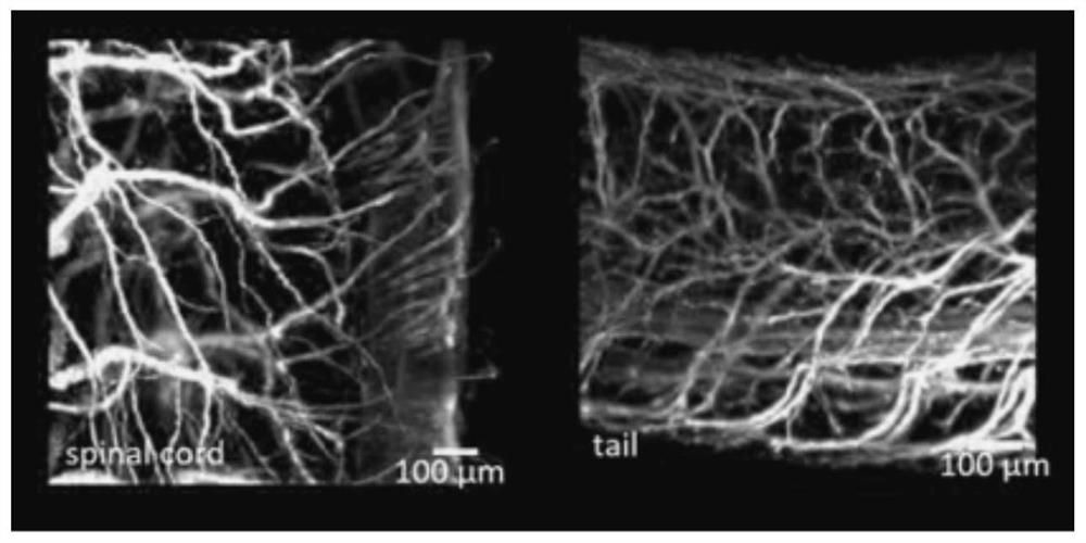
A tissue sample and immunofluorescence technology, applied in the biological field, can solve the problems of low image resolution, poor compatibility of fluorescent proteins, and low permeability of optical clearing agents, etc., to achieve clear neurons and blood vessels, and bright fluorescent markers.
- Summary
- Abstract
- Description
- Claims
- Application Information
AI Technical Summary
Problems solved by technology
Method used
Image

Examples
Embodiment 1
[0040] Sample Collection:
[0041] S1, first anesthetize the animal, and select the intraperitoneal injection of pentobarbital to anesthetize the mouse;
[0042] S2, the heart of the anesthetized mouse was perfused with PBS and 4% PFA sequentially;
[0043] S3, mice were dissected after perfusion, and mouse embryonic tissues were isolated;
[0044] S4, fix the embryo tissue with 4% PFA at 4°C, shake overnight, and then fix at 20-30°C for 1 hour;
[0045] S5, the embryo tissue was washed with PBS buffer at room temperature for 3 times, 30 minutes each time, and then the embryo tissue was placed in PBS buffer and stored at 4°C.
[0046] The fluorescent labeling method is as follows:
[0047] Transparent agent: the pH value is 7.5, including 2% urea by mass fraction, 20% DMSO by volume fraction, 2% sucrose by mass fraction, 3% SDS by mass fraction, 1% glutathione by mass fraction and 0.02% sodium azide and PBS buffer.
[0048] Blocking solution: PBS buffer, tritonx-100 and s...
Embodiment 2
[0064] Sample Collection:
[0065] S1, first anesthetize the animal, and select the intraperitoneal injection of pentobarbital to anesthetize the mouse;
[0066] S2, the heart of the anesthetized mouse was perfused with PBS and 4% PFA sequentially;
[0067] S3, the mouse was dissected after perfusion, and the brain tissue of the mouse was isolated;
[0068] S4, fix the brain tissue with 4% PFA at 4°C, shake overnight, and then fix at 20-30°C for 1 hour;
[0069] S5, the brain tissue was washed with PBS buffer solution at room temperature for 3 times, 30 minutes each time, and then the brain tissue was placed in PBS buffer solution and stored at 4°C.
[0070] The fluorescent labeling method is as follows:
[0071] Transparent agent: the pH value is 8.5, including the urea of 3% by mass fraction, the DMSO of 30% by volume fraction, the sucrose of 5% by mass fraction, the SDS of 5% by mass fraction, the glutathione and the glutathione of 3% by mass fraction 0.05% sodium azi...
Embodiment 3
[0087] Sample collection:
[0088] S1, first anesthetize the animal, and select the intraperitoneal injection of pentobarbital to anesthetize the mouse;
[0089] S2, the heart of the anesthetized mouse was perfused with PBS and 4% PFA sequentially;
[0090] S3, the mouse was dissected after perfusion, and the brain tissue of the mouse was isolated;
[0091] S4, fix the brain tissue with 4% PFA at 4°C, shake overnight, and then fix at 20-30°C for 1 hour;
[0092] S5, the brain tissue was washed with PBS buffer solution at room temperature for 3 times, 30 minutes each time, and then the brain tissue was placed in PBS buffer solution and stored at 4°C.
[0093] The fluorescent labeling method is as follows:
[0094] Transparent agent: the pH value is 8, including the urea of 2.5% by mass fraction, the DMSO of 25% by volume fraction, the sucrose of 3% by mass fraction, the SDS of 4% by mass fraction, the glutathione of 2% by mass fraction and the mass fraction of 0.03% sodiu...
PUM
 Abstract
Description
Claims
Application Information