Image processing method and image processing device

A technology for image processing and CT images, applied in the fields of image processing, computer-readable storage media, and image processing devices, to achieve the effects of reducing medical costs, avoiding trauma and potential risks, and avoiding radiation

Active Publication Date: 2021-06-22
INFERVISION MEDICAL TECH CO LTD
View PDF6 Cites 3 Cited by
  • Summary
  • Abstract
  • Description
  • Claims
  • Application Information

AI Technical Summary

Problems solved by technology

[0004] In view of this, the embodiment of the present application provides an image processing method, an image processing device, an electronic device, and a computer-r

Method used


Image

  • Image processing method and image processing device
  • Image processing method and image processing device
  • Image processing method and image processing device

Examples


Embodiment Construction

[0034] The following will clearly and completely describe the technical solutions in the embodiments of the application with reference to the drawings in the embodiments of the application. Apparently, the described embodiments are only some, not all, embodiments of the application. Based on the embodiments in this application, all other embodiments obtained by persons of ordinary skill in the art without creative efforts fall within the protection scope of this application.

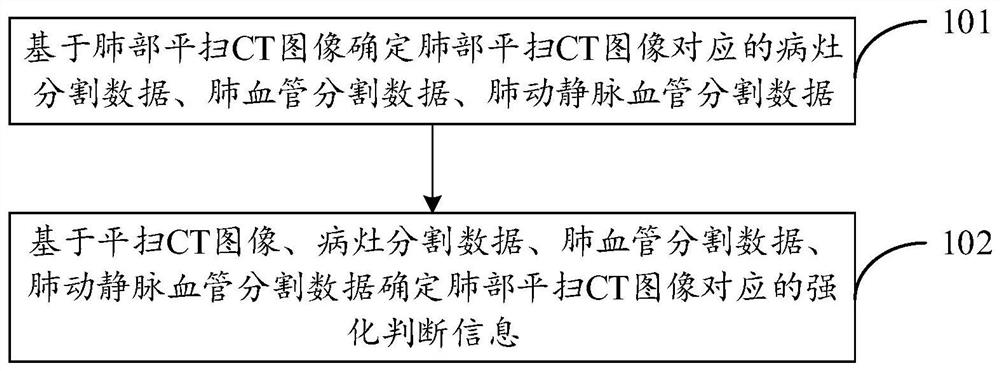
[0035] Computer tomography (Computed Tomography, CT) mainly uses precisely collimated X-ray beams, γ-rays and ultrasonic waves, etc., together with highly sensitive detectors to scan a certain part of the human body one by one, with a scan time Fast, clear images and other characteristics, can be used for the inspection of many diseases.

[0036] CT examination includes plain scan CT examination and enhanced CT examination. Ordinary CT examination performed directly on the instrument without contrast age...


PUM


Abstract

The invention provides an image processing method and an image processing device. The image processing method comprises the steps: determining focus segmentation data, lung blood vessel segmentation data and lung arteriovenous blood vessel segmentation data corresponding to a lung plain-scan CT image based on the lung plain-scan CT image; and determining reinforcement judgment information corresponding to the lung plain-scan CT image based on the plain-scan CT image, the focus segmentation data, the lung blood vessel segmentation data and the lung arteriovenous blood vessel segmentation data; and judging whether initial abnormal lesions exist in the lung or not by judging the strengthening state of the focus area. As the contrast agent can be prevented from being injected without enhanced CT examination, wounds and potential risks caused by injection of the contrast agent are avoided. Meanwhile, radiation to the patient during CT examination is prevented from being enhanced, and the doctor seeing cost of the patient is reduced.

Description

technical field [0001] The present application relates to the technical field of medical image processing, and in particular to an image processing method, an image processing device, electronic equipment, and a computer-readable storage medium. Background technique [0002] CT examination is an important means of screening cancer and other diseases, CT examination includes plain scan CT examination and enhanced CT examination. It is not easy to find abnormal lesions at the early stage of the lesion in plain CT examination, and enhanced CT examination is needed to judge the abnormal lesion in the early stage of the lesion through the enhancement of the lesion. [0003] However, enhanced CT examination needs to inject contrast agent into the patient, and the human body will have different degrees of adverse reactions to the contrast agent. How to avoid the use of contrast agents and accurately detect abnormal lesions in the early stage of lesions has attracted much attention...

Claims


Application Information

Patent Timeline
IPC IPC(8): G06T7/00G06T7/11
CPCG06T7/0012G06T7/11G06T2207/10081G06T2207/30061G06T2207/30096G06T2207/30101G06T2207/20081G06T2207/20084
Inventor 张荣国夏晨张华杰张欢王少康陈宽
Owner INFERVISION MEDICAL TECH CO LTD
Why Eureka
Try Eureka