Mouse-derived oral squamous cell carcinoma cell strain, and preparation method and application thereof
A technology of squamous cells and cancer cells, applied in the field of cell lines
- Summary
- Abstract
- Description
- Claims
- Application Information
AI Technical Summary
Problems solved by technology
Method used
Image
Examples
Embodiment 1
[0025] Embodiment 1 Preparation method of mouse oral squamous cell carcinoma cell line
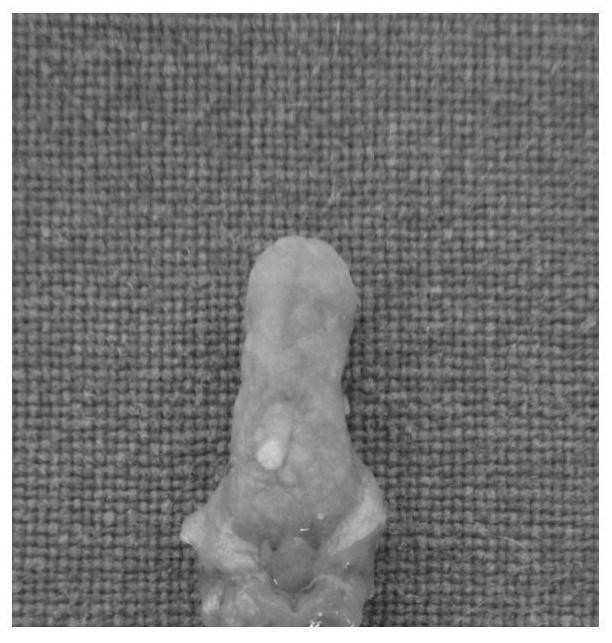
[0026] Step 1: Add 4-NQO (100 μg / ml) to the drinking water of C57BL / 6J mice for 20 weeks of induction, then replace with normal drinking water, and continue feeding for 4 weeks. A cauliflower-like lesion grows on the back of the mouse tongue. With invasion, the diagnosis was oral squamous cell carcinoma ( figure 1 ). Use high-temperature and high-pressure sterilized tissue scissors and blades to quickly excise the tumor tissue, soak it in high-sugar DMEM culture medium containing 20% fetal bovine serum and 1% penicillin + streptomycin double antibody, and quickly transfer it to a biological safety cabinet for operation . Wash repeatedly with PBS buffer containing 1% double antibody to obtain mouse tumor tissue;
[0027] Step 2: Cut the above mouse tumor tissue into 1*1*1mm small pieces with sterilized tissue scissors, wash with PBS, add 1 ml of trypsin, and incubate in a 37°C cell cul...
Embodiment 2
[0030] Example 2 Detection and identification of mouse oral squamous cell carcinoma cell lines
[0031] (1) Morphological observation
[0032] The cell shape is polygonal, oval or kidney-shaped, and grows in clusters, with large nuclei and abundant cytoplasm.
[0033] (2) Immunofluorescence staining detection
[0034] Step 1: Inoculate the cells on a cell slide, and when the cell density reaches 40% to 50%, rinse twice with PBS buffer for 5 minutes each time, add 4% paraformaldehyde to fix for 30 minutes, and then rinse again with PBS buffer. Rinse twice, 5 minutes each time;
[0035] Step 2: Add 100 microliters of 3% hydrogen peroxide solution to each slice, in order to block endogenous catalase and reduce non-specific staining, incubate at room temperature for 30 minutes, wash with PBS buffer 3 times, each 5 minute;
[0036] Step 3: Add 100 microliters of 5% goat serum to each tablet and incubate at room temperature for 20 minutes;
[0037] Step 4: Add 100 microliters o...
Embodiment 3
[0047] The establishment of the animal model of embodiment 3 mouse oral squamous cell carcinoma cell lines
[0048] Step 1: Anesthetize the C57BL / 6J mice, depilate the upper back, and prepare the above cells into 1*10 7 cells / 1 ml of cell suspension, each mouse was inoculated with 100 microliters of cells;
[0049] Step 2: Observe the state of the mice twice a week, measure the tumor size of the mice (tumor volume=maximum tumor diameter*minimum diameter*minimum diameter / 2), and weigh the body weight of the mice;
[0050] Step 3: When the tumor reaches 1000mm 2 The mice were euthanized before, and the tumor tissues of the mice were collected, fixed in formaldehyde, embedded in paraffin, and sectioned;
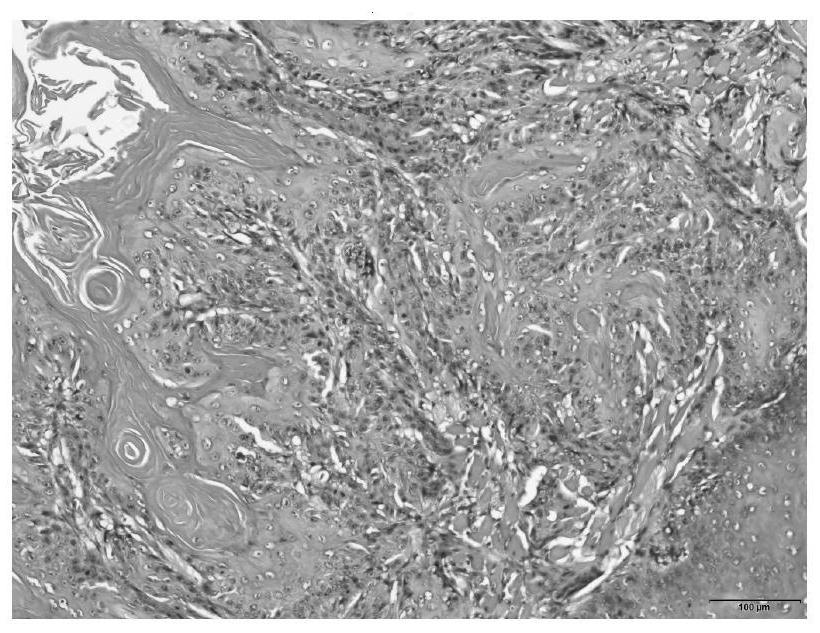
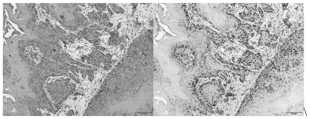
[0051] Step 4: Perform HE and immunofluorescence staining on the slices, see the results Figure 7 , Figure 8 .
[0052] To sum up, observed under an optical microscope, the cells were polygonal or elliptical, growing in clusters and adherent to the wall, with large nuclei a...
PUM
 Abstract
Description
Claims
Application Information
 - R&D
- Intellectual Property
- Life Sciences
- Materials
- Tech Scout
- Unparalleled Data Quality
- Higher Quality Content
- 60% Fewer Hallucinations
Browse by: Latest US Patents, China's latest patents, Technical Efficacy Thesaurus, Application Domain, Technology Topic, Popular Technical Reports.
© 2025 PatSnap. All rights reserved.Legal|Privacy policy|Modern Slavery Act Transparency Statement|Sitemap|About US| Contact US: help@patsnap.com


