A Bottom-Up Classification Method of Parasite Species Developmental Stages and Image Pixels
A developmental stage, bottom-up technology, applied in image analysis, image enhancement, image data processing, etc., can solve problems such as insurmountable long tail problem, contribution weight distinction, poor detection effect, etc., to reduce labor costs and regional Medical disparities, suppression numbers, effects of improving parasite detection
- Summary
- Abstract
- Description
- Claims
- Application Information
AI Technical Summary
Problems solved by technology
Method used
Image


Examples
Embodiment 1
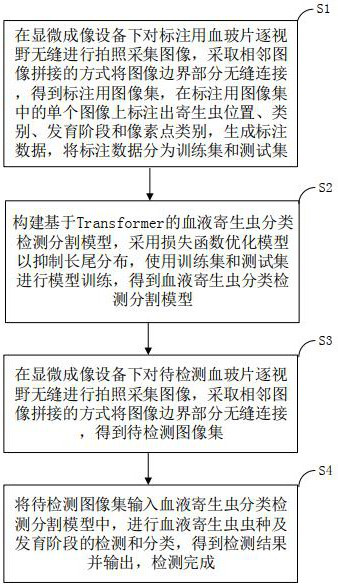
[0094] Such as figure 1 As shown, a bottom-up parasite species development stage and image pixel classification method includes the following steps:
[0095]S1. Preparation of training set and test set: under the microscope imaging equipment, the blood slides for labeling are seamlessly photographed and collected by field of view, and the border parts of the images are seamlessly connected by stitching adjacent images to obtain the image set for labeling , mark the position, category, developmental stage and pixel point category of the parasite on a single image in the image set for labeling, generate labeling data, and divide the labeling data into a training set and a test set;
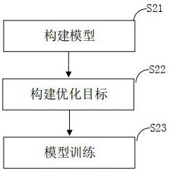
[0096] S2. Construction of Transformer-based blood parasite classification, detection and segmentation model: build a Transformer-based blood parasite classification, detection and segmentation model, use a loss function to optimize the model to suppress the long-tail distribution, use the training...
Embodiment 2
[0100] Such as figure 1 As shown, a bottom-up parasite species development stage and image pixel classification method includes the following steps:
[0101] S1. Preparation of training set and test set: under the microscope imaging equipment, the blood slides for labeling are seamlessly photographed and collected by field of view, and the border parts of the images are seamlessly connected by stitching adjacent images to obtain the image set for labeling , mark the position, category, developmental stage and pixel point category of the parasite on a single image in the image set for labeling, generate labeling data, and divide the labeling data into a training set and a test set;
[0102] S2. Construction of Transformer-based blood parasite classification, detection and segmentation model: build a Transformer-based blood parasite classification, detection and segmentation model, use a loss function to optimize the model to suppress the long-tail distribution, use the trainin...
Embodiment 3
[0133] Such as image 3 As shown, a bottom-up parasite species development stage and image pixel classification method includes the following steps:
[0134] 1. Whole film collection and splicing
[0135] In order to avoid the missed detection of parasites, the present invention first seamlessly takes pictures and collects the blood slides field by field under the microscopic imaging equipment, and for the image boundary, adopts the mode of adjacent image splicing to seamlessly connect the boundary parts, so as to avoid Leave out any possible parasites.
[0136] 2. Parasite data labeling
[0137] Professional doctors use specific labeling tools to mark the location of parasites, category information and pixel category information on a single field of view. After the amount of labeling reaches a certain scale, the labeling data is divided into a training set and a test set according to a certain ratio. Learn to prepare data for network models.
[0138] 3. Construction of Tr...
PUM
 Abstract
Description
Claims
Application Information
 - R&D
- Intellectual Property
- Life Sciences
- Materials
- Tech Scout
- Unparalleled Data Quality
- Higher Quality Content
- 60% Fewer Hallucinations
Browse by: Latest US Patents, China's latest patents, Technical Efficacy Thesaurus, Application Domain, Technology Topic, Popular Technical Reports.
© 2025 PatSnap. All rights reserved.Legal|Privacy policy|Modern Slavery Act Transparency Statement|Sitemap|About US| Contact US: help@patsnap.com