

Method for detecting environmental low-dose ionizing radiation
A technology for ionizing radiation and detection environment, applied in the field of radiation detection to achieve good sensitivity and specificity
- Summary
- Abstract
- Description
- Claims
- Application Information
AI Technical Summary
Problems solved by technology
Method used
Image

Examples
Embodiment 1
[0043] 1. Cell culture: The cell lines human lymphoblastoid AHH-1 and human bronchial epithelial cell 16HBE used in the present invention are cultured in 1640 medium and DMEM medium containing 10% fetal bovine serum respectively, and placed at 37° C. for 5 %CO 2 Subculture in an incubator.
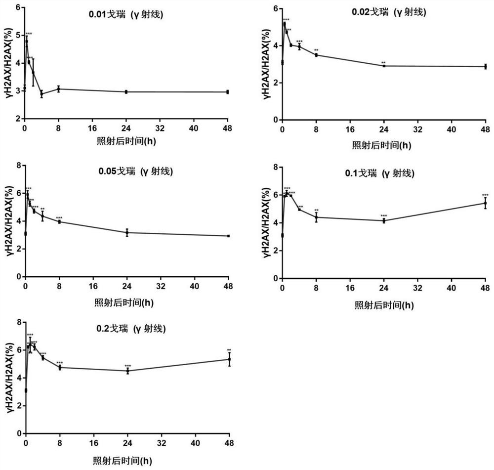
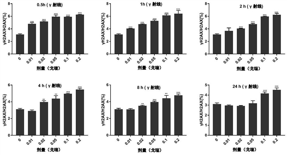
[0044] 2. Irradiation with cobalt-60γ-rays: Subcultured AHH-1 cells and 16HBE cells were irradiated with cobalt-60γ-rays. Wherein, the irradiation conditions of cobalt-60γ-ray irradiation are 20-25° C., the irradiation distance is 3 meters, and the dose rate is 2.81 cGy / min. The cells (marked as 0Gy) of the control group (non-irradiated group) were collected, washed with phosphate buffered saline in turn, and the cells of 0.5 hour, 1 hour, 2 hours, 4 hours, 8 hours, 24 hours and 48 hours after irradiation were collected ( Marked as 0.5h, 1h, 2h, 4h, 8h, 24h and 48h). The irradiation doses were 0.01Gy, 0.02Gy, 0.05Gy, 0.1Gy, 0.2Gy, respectively.
[0045] 3. Extraction of nuclear protein...
Embodiment 2
[0089] 1. The peripheral blood (3mL) of 16 females and 25 males was extracted to extract lymphocytes for the detection of the background value of the negative control group.
[0090] The present invention is based on the steps of extracting lymphocytes from human peripheral blood lymphocyte separation fluid (Tianjin Hanyang); this specification requires extracting lymphocytes at room temperature, but the lymphocytes extracted at room temperature in the present invention are detected by the method of the present invention , showing no signal, that is, no γH2AX can be detected. However, when the lymphocytes were extracted at 4°C for detection, a weaker signal could be seen. Therefore, the present invention chooses to extract lymphocytes at 4°C. At the same time, because the rotational speed setting in the manual of human peripheral blood lymphocyte separation fluid is designed under normal temperature conditions, it is not suitable for extraction at low temperature. The present...
Embodiment 3
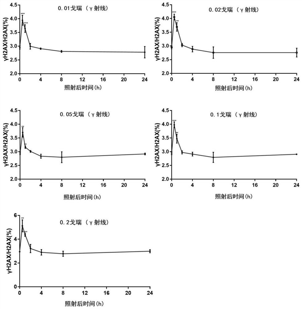
[0104] The fitting curve was constructed on the level of human peripheral blood lymphocytes. That is, the value of γH2AX / H2AX (%) after 0.01Gy, 0.02Gy, 0.05Gy, 0.1Gy and 0.2Gy low-dose irradiation of human peripheral blood for 0.5h is the ordinate, and the radiation dose (0.01Gy, 0.02Gy, 0.05Gy, 0.1Gy and 0.2Gy) are constructed as the abscissa, the result is as follows Figure 10 shown. The result shows: the fitting equation is y=-104.99x 2 +37.689x+3,0409,R 2 =0.9632, indicating that the fitting degree between the estimated value of the fitting curve and the corresponding actual data is high, which can be used to evaluate the received radiation dose.
PUM
 Abstract
Description
Claims
Application Information