Prussian blue nano MRI tracer agent as well as preparation method and application thereof
A Prussian blue and tracer technology, applied in the field of Prussian blue nano-MRI tracer and its preparation, achieves the effects of long-distance transportation, good biocompatibility, biosafety, and good stability
- Summary
- Abstract
- Description
- Claims
- Application Information
AI Technical Summary
Problems solved by technology
Method used
Image
Examples
Embodiment 1
[0035] 1) Preparation of cysteine modified β-amyloid 42 (cys-Aβ42)
[0036] A cysteine containing a sulfhydryl group is inserted between the 28th lysine and the 29th glycine of β-amyloid 42 to synthesize cysteine-modified β-amyloid 42, whose sequence is: DAEFRHDSGYEVHHQKLVFFAEDVGSNK(Cys)GAIIGLMVGGVVIA.
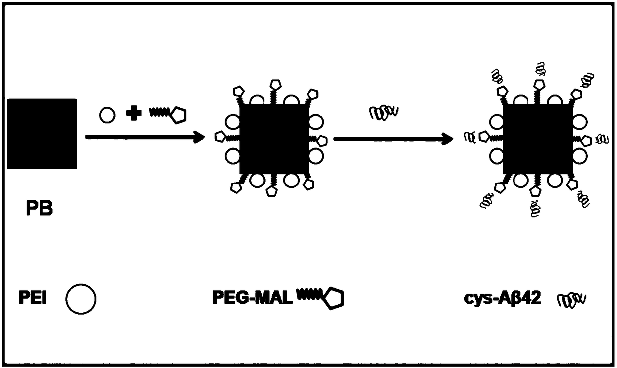
[0037] 2) Preparation of maleimidized Prussian blue nanoparticles (PB@PEI@MAL NPs)
[0038] Dissolve 0.5mM citric acid in 1mM FeCl3·6H2O deionized water solution to prepare mixture A, and dissolve 0.5mM citric acid in 1mM K4Fe(CN)6·3H2O deionized water solution to prepare mixture B, and then heat it at 60℃ Add 20ml of mixed solution B dropwise to 20ml of mixed solution A, and continue to stir for 5 minutes, the mixed solution will turn bright dark blue. Add a polyetherimide (PEI) solution with a concentration of 0.005mM, keep stirring at 60°C for 4 hours, centrifuge at 15000rpm for 30min to collect the precipitate and wash with acetone and deionized water, and finally resuspend...
Embodiment 2
[0044] The Prussian blue nano-NMR tracer PB@PEI@MALcys-Aβ42 NPs prepared in Example 1 was detected and characterized.
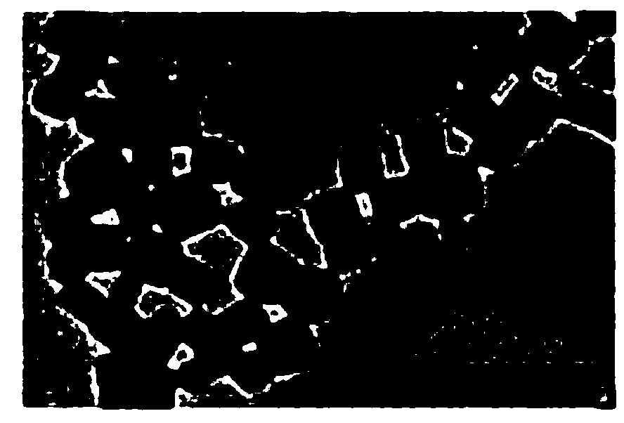
[0045] figure 2 It is a transmission electron micrograph of the Prussian blue nano-NMR tracer PB@PEI@MAL-cys-Aβ42 NPs prepared in Example 1.
[0046] The morphology of PB@PEI@MAL-cys-Aβ42 NPs was characterized by transmission electron microscope, the specific operation steps are as follows:
[0047] Dissolve PB@PEI@MAL-cys-Aβ42 NPs in PBS and then disperse ultrasonically. Pipette 10μl of the solution onto a copper mesh covered with ultra-thin carbon film, and fully adsorb for 10-30 minutes until the sample is dry and then proceed in a transmission electron microscope. Observe and take pictures. The result is figure 2 As shown, it can be seen that the particle size of PB@PEI@MAL-cys-Aβ42 NPs is about 60 nm, with a cubic structure, with a relatively uniform particle size distribution and good dispersion.
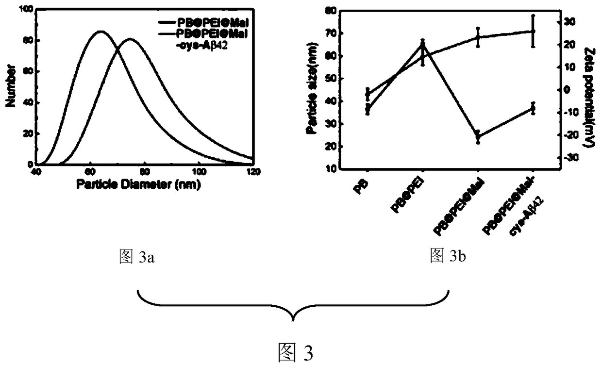
[0048] image 3 It is the particle size diagram of the Pru...
Embodiment 3
[0056] Figure 5 It is a graph of the relaxation time T1 and T2 with concentration in the MRI of the Prussian blue nano-NMR tracer PB@PEI@MAL-cys-Aβ42 NPs prepared in Example 1.
[0057] Configure PB@PEI@MAL-cys-Aβ42 NPs solution with a concentration gradient of 0-16mg / ml with PBS and place it in a NMR sample tube to evenly disperse; measure the spin of each concentration gradient solution by a 1.5 T NMR analyzer- Lattice relaxation time (T1) and spin-spin relaxation time (T2), while taking grayscale images. Figure 5 The relaxation time T1 and T2 of the weighted NMR imaging of the Prussian blue nano-NMR tracer PB@PEI@MAL-cys-Aβ42 NPs increased with the increase of the concentration of PB@PEI@MAL-cys-Aβ42NPs .
PUM
| Property | Measurement | Unit |
|---|---|---|
| Particle size | aaaaa | aaaaa |
| Particle size | aaaaa | aaaaa |
| Particle size | aaaaa | aaaaa |
Abstract
Description
Claims
Application Information
 - R&D
- Intellectual Property
- Life Sciences
- Materials
- Tech Scout
- Unparalleled Data Quality
- Higher Quality Content
- 60% Fewer Hallucinations
Browse by: Latest US Patents, China's latest patents, Technical Efficacy Thesaurus, Application Domain, Technology Topic, Popular Technical Reports.
© 2025 PatSnap. All rights reserved.Legal|Privacy policy|Modern Slavery Act Transparency Statement|Sitemap|About US| Contact US: help@patsnap.com


