Monoclonal antibody for detecting mouse interleukin 6 as well as preparation method and application thereof
A monoclonal antibody and interleukin technology, applied in the field of immunoassay, can solve problems such as narrow linear range, low detection sensitivity, and radioactive contamination, and achieve the effects of wide linear range, good reaction specificity, and improved sensitivity and precision
- Summary
- Abstract
- Description
- Claims
- Application Information
AI Technical Summary
Problems solved by technology
Method used
Image


Examples
Embodiment 1
[0060] In this example, the monoclonal antibody for detecting mouse interleukin 6 comprises a first antibody and a second antibody, the first antibody is secreted by the hybridoma cell line 20-A4-D5, and the second antibody is secreted by the hybridoma cell Secreted by strain 10-B8-C9, the hybridoma cell strain 20-A4-D5 and the hybridoma cell strain 10-B8-C9 were prepared by the following method:
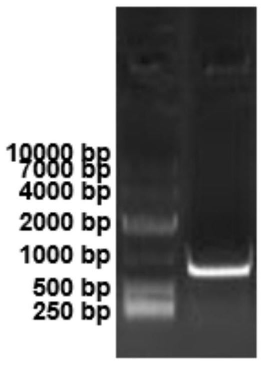
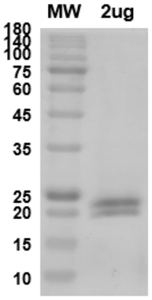
[0061] S1. Artificially synthesizing the mouse interleukin-6 gene, recombining the mouse interleukin-6 gene into the expression vector plasmid pATX1 to obtain the IL6-pATX1 expression vector, and verifying the vector by electrophoresis, the results are as follows figure 1 shown by figure 1 It can be seen that the cloning site is EcoRI / XhoI, and the sequence of the mouse interleukin 6 gene is shown in SEQ ID No.13; the IL6-pATX1 expression vector was transfected into the 293F cell line and cultivated for 6 days, and the supernatant was collected The solution was purified by nickel c...
PUM
 Abstract
Description
Claims
Application Information