Application of M2 type macrophage exosome in preparation of medicine for treating osteoporosis
A technology for osteoporosis and macrophages, applied in the biological field, can solve problems such as application limitations, and achieve the effect of inhibiting bone loss.
- Summary
- Abstract
- Description
- Claims
- Application Information
AI Technical Summary
Problems solved by technology
Method used
Image
Examples
Embodiment 1
[0019] Example 1. Isolation and identification of M2 macrophage exosomes
[0020] Induction of S1.M2 macrophages
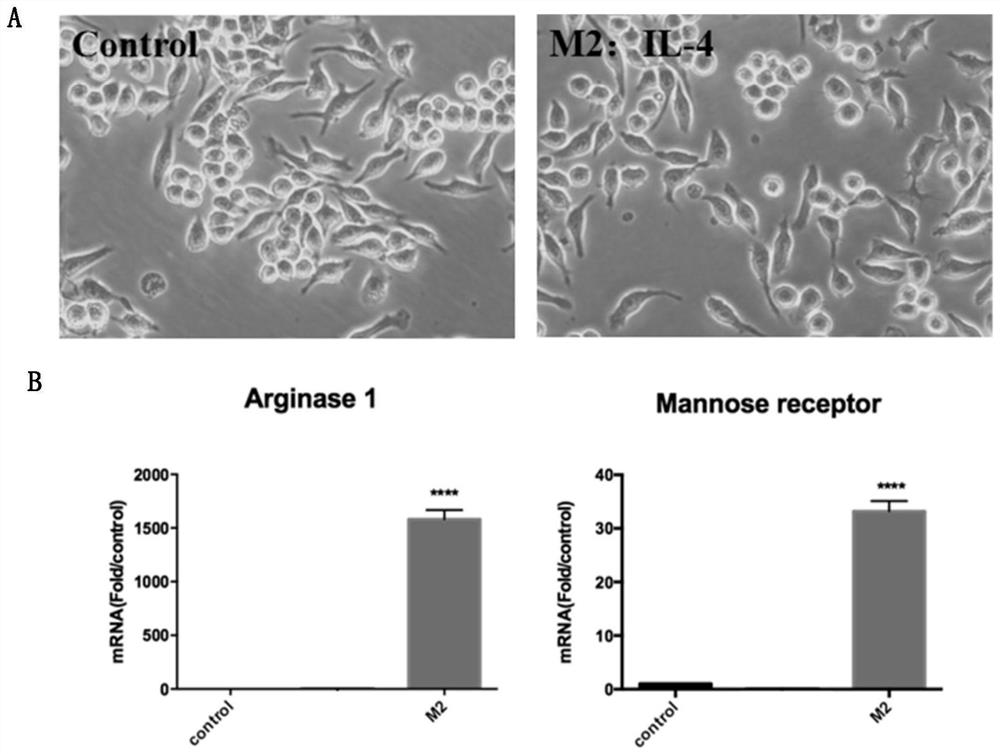
[0021] RAW264.7 (mouse mononuclear macrophage leukemia cells) was cultured in a 10cm dish containing 10% fetal calf serum in DMEM high-glucose medium, and when the cell proliferation was close to 70-80% confluent, it was passed on to a 10cm dish, The culture medium is DMEM high-glucose medium without fetal bovine serum, stimulated with 10ng / ml IL-4 every other day, and induced for 24 hours. figure 1 Display: After IL-4 stimulation, the cell morphology is "spindle-shaped", and RT-qPCR detection shows that the cells can express the classic markers of M2 macrophages: arginine (Arg-1) and mannitol receptor (Mannose receptor) . After confirming that the induced cells were M2 macrophages, the supernatant of the cells was collected for subsequent experiments.
[0022] S2. M2-EVs isolation
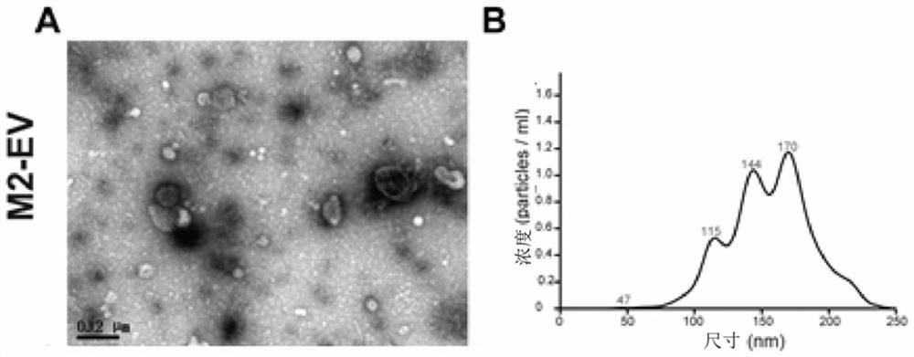
[0023] The supernatant collected above was subjected to exosome extraction. ...
Embodiment 2
[0029] Example 2, the inhibitory effect of M2-EVs on osteoclasts
[0030] S1. Isolation of mouse bone marrow-derived macrophages (BMM)
[0031] Mouse bone marrow-derived macrophages (BMM) were isolated according to conventional methods. After isolation, the BMMs were cultured in a 10 cm dish containing 10% fetal calf serum, 30ng / ml MCSF, and alpha MEM high-glucose medium containing double antibodies to penicillin and streptomycin , the medium was changed every 2 days, and on the 5th day of induction, the cells could proliferate to 70% confluence, and the cells were collected for subsequent experiments.
[0032] S2. Induction of osteoclasts
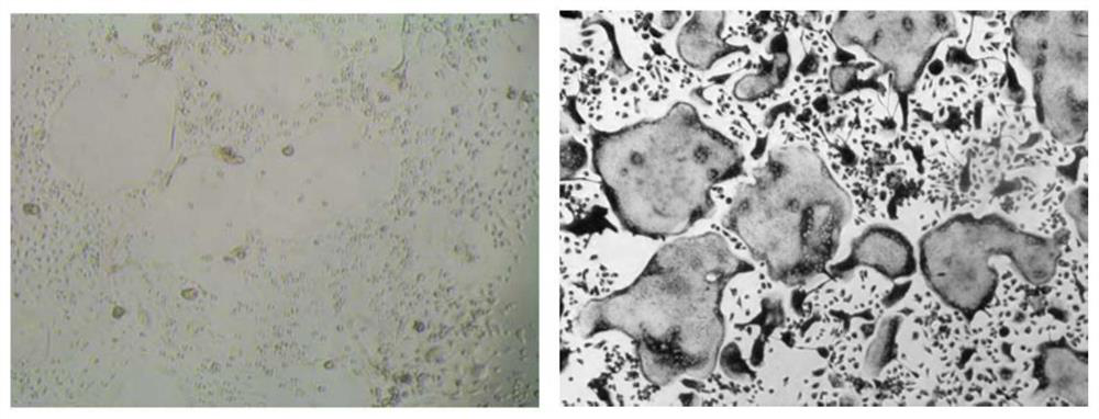
[0033] The above-mentioned cells were digested and seeded in a 96-well plate, and 100,00 BMMs were plated in each well. The medium was the same as above, and RANKL was added every other day to a final concentration of 100ng / ml, and the medium was changed every two days. image 3 It was shown that after induction, the cells began to fuse ...
Embodiment 3
[0037] Example 3, the rescue effect of M2-EVs on osteoporotic mice
[0038] Ovariectomized (OVX) mice were obtained by conventional ovariectomy in female mice of 8 weeks, and 1 μg / ul M2-EVs 100 μl (solvent was 10% PBS buffer) was injected through the tail vein after 1 week, and injected weekly 2 times. Eight weeks after injection, micro CT was used to analyze the femoral bone formation of OVX mice injected with M2-EVs regularly and the vehicle-injected control group (phosphate-buffered saline solution). Figure 5 The results showed that the addition of M2-EVs could effectively improve the bone loss in OVX mice.
PUM
 Abstract
Description
Claims
Application Information
 - R&D
- Intellectual Property
- Life Sciences
- Materials
- Tech Scout
- Unparalleled Data Quality
- Higher Quality Content
- 60% Fewer Hallucinations
Browse by: Latest US Patents, China's latest patents, Technical Efficacy Thesaurus, Application Domain, Technology Topic, Popular Technical Reports.
© 2025 PatSnap. All rights reserved.Legal|Privacy policy|Modern Slavery Act Transparency Statement|Sitemap|About US| Contact US: help@patsnap.com


