
Multi-thread strain imaging method and device based on living body ultrasonic image

An ultrasonic image and imaging method technology, applied in the field of medical image processing, can solve the problems of low time density, poor real-time performance, and large errors of output results, and achieve the effects of improving signal-to-noise ratio, improving accuracy, and efficient data processing

Pending Publication Date: 2022-03-25
NANJING UNIV
View PDF2 Cites 1 Cited by
  • Summary
  • Abstract
  • Description
  • Claims
  • Application Information

AI Technical Summary

Problems solved by technology

[0008] Aiming at the motion suppression realized by auxiliary gating of additional equipment in the existing technology, and because this kind of gating technology relies on a single motion phase, the output result has low time density and poor real-time performance, often accompanied by artifacts and large errors. The present invention provides a multi-threaded strain imaging method and device based on in vivo ultrasound images, which can obtain strain distribution in multiple motion phases, and further combine the spatial correlation denoising and fusion of strain distribution to obtain accurate and high robustness , In vivo strain distribution image with high time density

Method used


Image

  • Multi-thread strain imaging method and device based on living body ultrasonic image
  • Multi-thread strain imaging method and device based on living body ultrasonic image
  • Multi-thread strain imaging method and device based on living body ultrasonic image

Examples


Embodiment 1

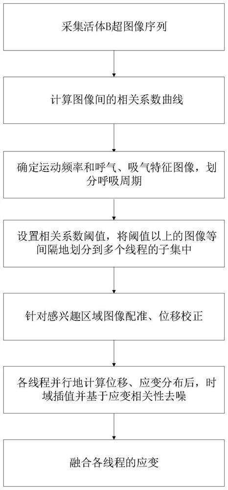
[0066] combine figure 1 , a kind of multi-threaded strain imaging method based on in vivo ultrasonic image of the present invention, comprises the following steps:

[0067] Acquisition step: carry out ultrasonic image acquisition to living object, obtain digital ultrasonic image sequence I:

[0068] For living objects in a state of regular physiological movement, B-mode ultrasound imaging is performed on the biological tissue in the target area to be inspected, and data is collected at equal time intervals to obtain digital image sequence I;

[0069] Specifically, the B-ultrasound instrument works in real-time imaging mode, adjusts the position, angle and imaging parameters of the ultrasonic probe to ensure that the target tissue is clearly visible in the image, and the probe and system parameters are fixed; Displacement and strain distribution are formed; B-ultrasound collects digital images continuously and at equal time intervals; the collection time is not shorter than 2 ...

Embodiment 2

[0113] This embodiment adopts the method in the foregoing embodiment 1, heats the visceral fat of living pigs with a microwave ablation needle and collects B-ultrasound digital image signals, and calculates the thermal strain based on the B-ultrasound images in vivo multi-threaded strain imaging method. The specific steps are as follows :

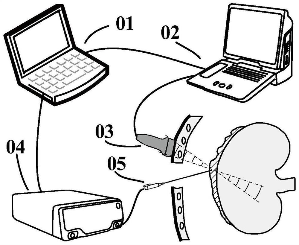
[0114] A multi-threaded strain imaging device based on in vivo ultrasound images such as figure 2 As shown, the digital ultrasonic image acquisition of the portable B-ultrasound instrument 02 is controlled by the computer 01. The digital sampling rate of the B-ultrasound instrument is 40MHz, and the imaging probe used is a linear array probe 03 with 128 array elements, and its center frequency is 10.5MHz. Under the guidance of the real-time B-ultrasound image presented by the B-ultrasound instrument, adjust the imaging position and angle of the ultrasound probe and the parameters of the imaging system, so that the target tissue is clearly ...


PUM


Abstract

The invention discloses a multi-thread strain imaging method and device based on a living body ultrasonic image, and belongs to the field of medical image processing. The method comprises the following steps: collecting a living body B ultrasonic image sequence, analyzing the correlation characteristics of the image, judging the respiratory rate, and dividing the image into different respiratory cycles and synchronous phase periods during the cycles; a calculation thread is established for each phase period, after displacement correction and image registration, the threads calculate strain in parallel, and noise reduction is carried out according to correlation between the threads; and finally, fusing multi-thread strain results to obtain accurate and high-time-density strain output. According to the method, the problems of artifacts and errors generated by physiological movement in strain imaging in the prior art can be solved, the strain distribution of multiple movement phase periods can be obtained, and the living body strain distribution image which is accurate, high in robustness and high in time density is obtained after denoising and fusion are further combined with the spatial correlation of the strain distribution.

Description

technical field [0001] The present invention relates to the field of medical image processing, more specifically, to a multi-thread strain imaging method and device based on ultrasound images of living bodies. Background technique [0002] Ultrasound imaging is one of the four major imaging diagnostic technologies in clinical medicine. It has high clinical application value due to its advantages of safety and non-invasiveness, low patient burden, and good real-time signal processing. The development of ultrasonic imaging technology has gone through from one-dimensional to three-dimensional, static to dynamic, structural imaging to functional imaging. The functions of ultrasonic diagnostic equipment are gradually enhanced, and the information provided is becoming more and more abundant. [0003] Along with the development of ultrasonic imaging technology and ultrasonic imaging equipment, a variety of diagnostic and analytical imaging algorithms based on ultrasonic imaging sig...

Claims


Application Information

Patent Timeline
IPC IPC(8): G06T5/50G06T5/00G06T7/38G06V10/25G06V10/44G06F9/50
CPCG06T5/50G06T7/38G06F9/5061G06T2207/10016G06T2207/10132G06T5/70
Inventor 郭霞生尹楚豪章东屠娟
Owner NANJING UNIV
Learn More