Bionic construction method of periosteum-like tissue
A construction method and technology of membrane tissue, applied in the intersecting fields of materials, life and medicine, can solve problems such as failure to meet critical defect repair needs, complex fabrication, low biological activity, etc.
- Summary
- Abstract
- Description
- Claims
- Application Information
AI Technical Summary
Problems solved by technology
Method used
Image
Examples
Embodiment 1
[0065] Preparation of implant material
[0066] Add 10 μg of recombinant human bone morphogenetic protein-7 (rhBMP-7) synthesized by eukaryotic or prokaryotic expression system to gelatin sponge (5 mm diameter × 5 mm thick, 10 mg weight), and freeze-dry to form an active material containing BMP-7.
[0067] Add 30 μg of recombinant human bone morphogenetic protein-2 (rhBMP-2) synthesized by eukaryotic or prokaryotic expression system into gelatin sponge (5mm diameter×5mm thickness, 10mg weight), and freeze-dry to form the active material containing BMP-2.
[0068] Add 30 μg rhBMP-2 and 100ng vascular endothelial growth factor (VEGF) synthesized by eukaryotic or prokaryotic expression system to gelatin sponge (5mm diameter×5mm thickness, 10mg weight), and freeze-dry to form the active material containing BMP-2 / VEGF .
[0069] 30 μg of rhBMP-2 synthesized by eukaryotic or prokaryotic expression system and 30 μg of chondroitin sulfate (CS) were added to gelatin sponge (5mm diamet...
Embodiment 2
[0071] In vivo construction of periosteoid tissue in mice
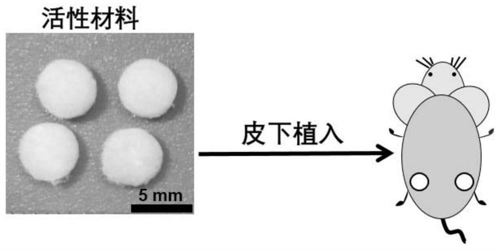
[0072] The periostoid tissue was formed in mice using the active material containing BMP-7 or BMP-2 or BMP-2 / VEGF or BMP-2 / CS described in Example 1. Such as figure 1 As indicated, the above materials were implanted subcutaneously in the back of 8-week-old C57BL / 6 male mice. The active material formed after implantation in mice developed into periosteoid tissue after 1 week. After feeding for 1 week, the constructed periostoid tissue was taken out, part of which was used to take macroscopic photos, tissue sections, etc. for the characterization of periostoid tissue, and the other part was used for autologous skull defect transplantation.
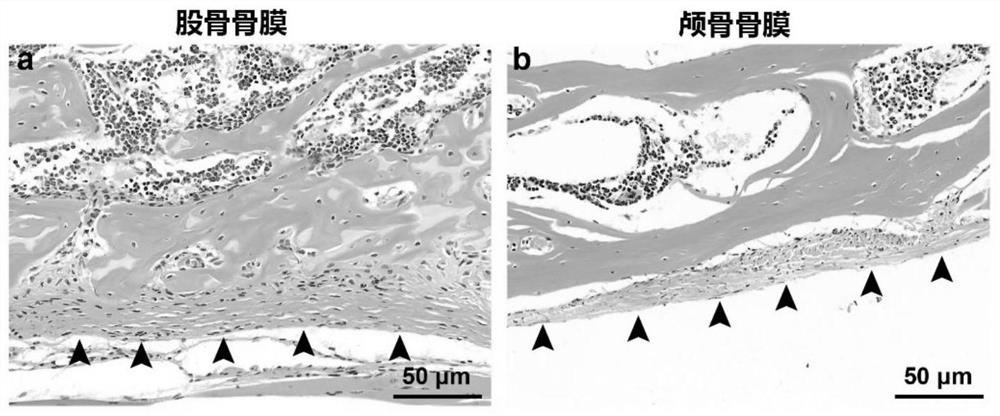
[0073] figure 2 H&E section of the periosteum tissue surrounding the native femur and skull, showing the microstructure of the femur and lateral skull in situ.
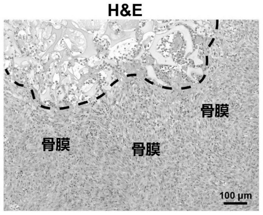
[0074] image 3 The shown hematoxylin / eosin (H&E) stained sections showed that: the periosteoid tissue formed in mi...
Embodiment 3
[0079] In vivo construction of periosteoid tissue for autologous calvarial defect therapy in mice
[0080] The purpose of this example is to evaluate the therapeutic effect of periostoid tissue produced in mice on autologous calvarial defects with a diameter of 5 mm.
[0081] grouping:
[0082] SPF grade C57BL / 6 mice, male, 8 weeks old, were randomly divided into groups. The experimental groups are as follows:
[0083] group blank group BMP-2 / CS group quantity 6 6
[0084] Preparation of periostoid tissue: The scaffold containing BMP-2 / CS described in Example 1 was implanted subcutaneously, and the periostoid tissue was produced after 1 week of development. The periostoid tissue was removed, and the resulting disc-shaped periostoid tissue with a diameter of 5 mm was trimmed using a 5 mm inner diameter punch.
[0085] Autologous periosteoid tissue transplantation: After the mice were anesthetized, the skin of the mouse head was cut open with a scalp...
PUM
| Property | Measurement | Unit |
|---|---|---|
| diameter | aaaaa | aaaaa |
Abstract
Description
Claims
Application Information
 - R&D
- Intellectual Property
- Life Sciences
- Materials
- Tech Scout
- Unparalleled Data Quality
- Higher Quality Content
- 60% Fewer Hallucinations
Browse by: Latest US Patents, China's latest patents, Technical Efficacy Thesaurus, Application Domain, Technology Topic, Popular Technical Reports.
© 2025 PatSnap. All rights reserved.Legal|Privacy policy|Modern Slavery Act Transparency Statement|Sitemap|About US| Contact US: help@patsnap.com


