Three-phase bionic sleeve stent and preparation method thereof

A sleeve stent and three-phase technology, applied in the field of medical devices, can solve problems such as shortage, poor healing of biocompatible materials and bone tunnels, and lack of grafts to induce stem cell differentiation, so as to achieve small cell impact and accelerate the bone tunnel. Regeneration of tendon insertion structure and reduction of decellularization time

Active Publication Date: 2022-04-12
XIANGYA HOSPITAL CENT SOUTH UNIV
View PDF7 Cites 0 Cited by
  • Summary
  • Abstract
  • Description
  • Claims
  • Application Information

AI Technical Summary

Problems solved by technology

At present, a variety of ACL reconstruction grafts such as allogeneic tendons and polymer artificial ligaments have been used clinically, but there are the following problems: 1) Most of the existing grafts have a single-phase structure imitating tendon tissue, and there is a lack of bone-fibrocartilage in the bone tunnel -The three-phase structure of tendon cannot provide supporting materials for the regeneration of bone-fibrocartilage-tendon three-phase structure in the bone canal; 2) The existing grafts lack the functional activity of inducing stem cells to differentiate into osteogenesis, cartilage and tendon, which is not conducive to osteogenesis The formation of new bone-tendon insertions in the tunnel and the regeneration of tendons in the joint cavity; 3) The grafts made of artificial synthetic materials have many problems such as degradation in vivo, biocompatibility, poor healing of materials and bone tunnels, etc.

Method used


Image

  • Three-phase bionic sleeve stent and preparation method thereof
  • Three-phase bionic sleeve stent and preparation method thereof
  • Three-phase bionic sleeve stent and preparation method thereof

Examples


Embodiment 1

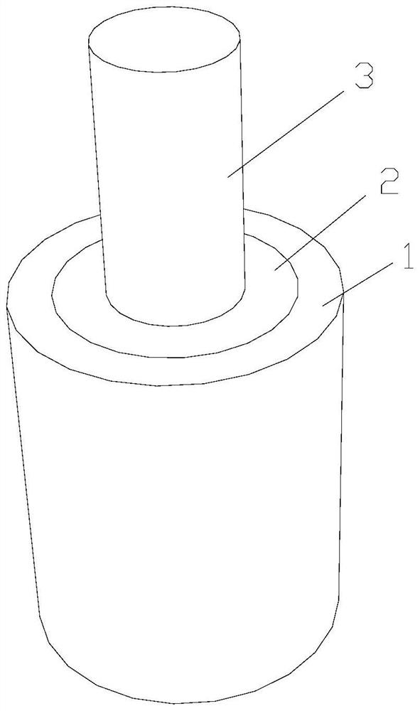
[0068] A kind of three-phase bionic sleeve support of the present invention, as figure 1 As shown, including bone sleeve support 1, cartilage sleeve support 2 and tendon support 3, bone sleeve support 1 and cartilage sleeve support 2 are hollow cylindrical structures, tendon support 3 is a cylindrical structure, tendon support 3 The cartilage sleeve support 2 is set on the outside of the cartilage sleeve support 2, and the bone sleeve support 1 is covered with the cartilage sleeve support 2, and the space between the bone sleeve support 1 and the cartilage sleeve support 2, and between the cartilage sleeve support 2 and the tendon support 3 are all For a tight fit.

[0069] As an implementation of this embodiment, the bone sleeve support 1 is made of decellularized bone, the cartilage sleeve support 2 is made of decellularized cartilage, and the tendon support 3 is made of decellularized tendon. The bone sleeve support is a homogeneous mineralized bone sleeve support, and the...


PUM

PropertyMeasurementUnit
lengthaaaaaaaaaa
diameteraaaaaaaaaa
lengthaaaaaaaaaa

Abstract

The invention discloses a three-phase bionic sleeve stent which comprises a bone sleeve stent, a cartilage sleeve stent and a tendon stent, the tendon stent is sleeved with the cartilage sleeve stent, the cartilage sleeve stent is sleeved with the bone sleeve stent, the bone sleeve stent is in close fit with the cartilage sleeve stent, and the cartilage sleeve stent is in close fit with the tendon stent. The preparation method comprises the following steps: preparing a bone sleeve scaffold, a cartilage sleeve scaffold and a tendon scaffold, carrying out decellularization treatment on each scaffold, constructing a C-BMP-2 modified decellularized bone sleeve scaffold, a C-TGF-beta3 modified decellularized cartilage sleeve scaffold and a C-GDF-7 modified decellularized tendon scaffold, and carrying out telescoping combination to obtain the product. The invention has the functional activity of inducing osteogenesis, cartilage formation and tendon differentiation of stem cells, can accelerate regeneration of tendon dead center structures in bone tunnels and tendon remodeling in articular cavities after ACL reconstruction, and realizes early joint function recovery after ACL reconstruction.

Description

technical field [0001] The invention belongs to the technical field of medical devices, and in particular relates to a three-phase bionic sleeve bracket and a preparation method thereof. Background technique [0002] The knee joint plays an important role in human activities. Among the ligaments that make up the knee joint, the anterior cruciate ligament (ACL) connects the femur and tibia of the knee joint, restricts the excessive forward movement of the tibia during knee flexion and extension, and is the most important ligament to maintain the stability of the knee joint. Unfortunately, ACL injury will lead to multidirectional instability of the knee joint, causing excessive stress on the cartilage articular surface and meniscus, accelerating its wear and tear, and eventually leading to osteoarthritis (OA). According to reports, ACL ruptures are common in sports involving twisting, half-bending, emergency stops, and collisions, such as football, basketball, badminton, and ...

Claims


Application Information

Patent Timeline
Patent Type & Authority Applications(China)
IPC IPC(8): A61L27/36A61L27/22A61L27/50A61L27/54A61F2/08
Inventor 吕红斌史强胡建中陈灿许琰段嵘杰寻金瑞
Owner XIANGYA HOSPITAL CENT SOUTH UNIV
Why Eureka
Try Eureka