Kit for simultaneously detecting lung cancer and pulmonary infection

A kit and reagent technology, applied in the biological field

Pending Publication Date: 2022-05-24
SUZHOU HONGYUAN BIOTECH CO LTD
View PDF0 Cites 0 Cited by
  • Summary
  • Abstract
  • Description
  • Claims
  • Application Information

AI Technical Summary

Problems solved by technology

[0006] There is no next-generation sequencing kit for simultaneous detection of lung infection and lung cancer in the prior art

Method used


Image

  • Kit for simultaneously detecting lung cancer and pulmonary infection
  • Kit for simultaneously detecting lung cancer and pulmonary infection
  • Kit for simultaneously detecting lung cancer and pulmonary infection

Examples


Embodiment 1

[0058] Example 1 alveolar lavage solution human and microbial genomic DNA extraction

[0059] The kit selected in the present embodiment was purchased from QIAGEN Corporation (item no.: 80204).

[0060]a. Transfer the alveolar lavage solution to a 50 mL centrifuge tube, add the same volume of 1× PBS buffer, shake and mix well, perform a 1600 g centrifugation for 10 min, carefully pour out the supernatant, and then carefully aspirate the remaining supernatant with a pipette.

[0061] B. Add 350 μL of 1× Buffer RL / DTT to a new 1.5 mL centrifuge tube and carefully whip the lysate at least 5 times with a disposable 20 gauge needle syringe to break up the cells.

[0062] c. Pack the DNA purification column in a 2 mL collection tube. Transfer the cell lysate to the gDNA filtration column. Centrifuge at 14,000 g for 2 min.

[0063] d. Take the DNA purification column and pack it in a 2 mL collection tube. Add 500 μL buffer DW1 to the column and let stand for 2 min. Centrifuge 10000 g for...

Embodiment 2

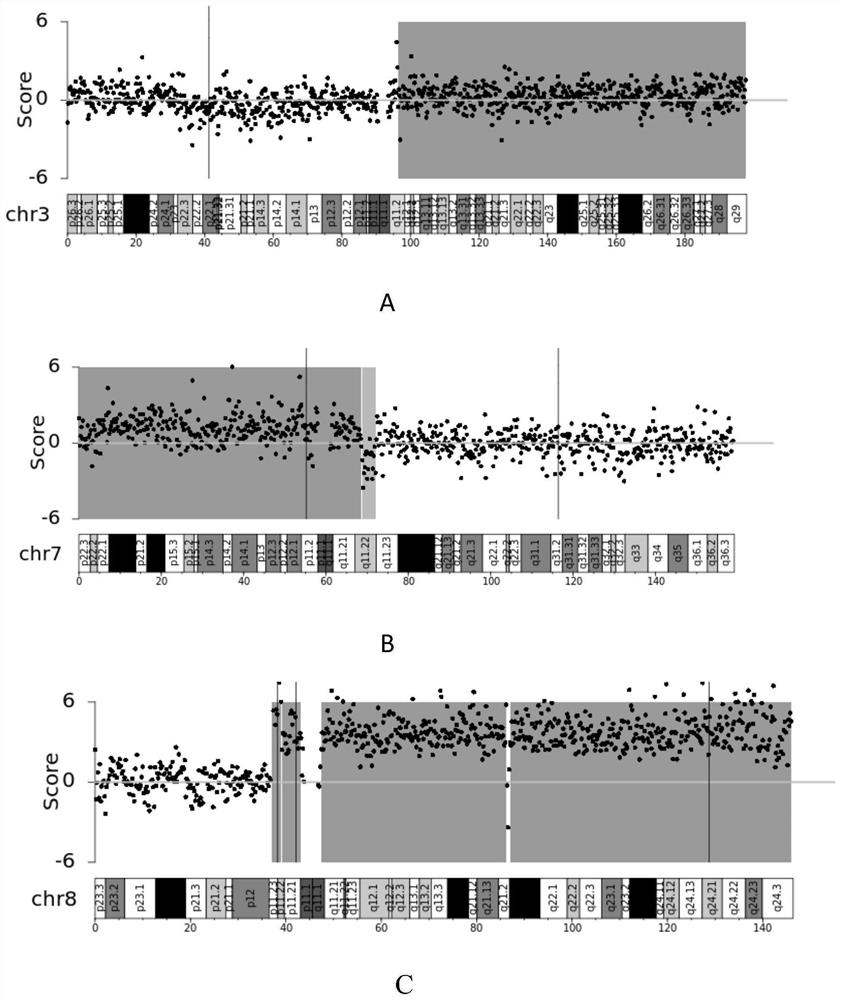
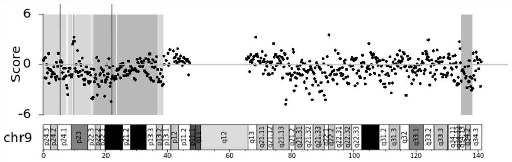
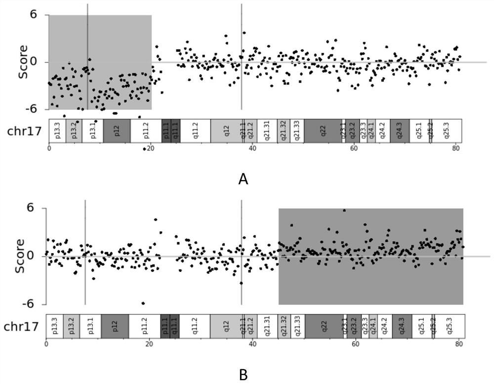
[0069] Example 2 Detection of chromosomal unstable regions and pathogenic microbial DNA

[0070] In the present embodiment, the relevant library kit was purchased from NEB Company, item number: E7645S;

[0071] These include the following steps:

[0072] 1. Genome fragmentation: take the 20ng human / microbial genomic DNA prepared in Example 1, prepare the digestion reaction system as shown in Table 1, and react according to the procedure of Table 2. In the present embodiment, the concentration of human genomic DNA is 2ng / μL, and 10 μL is added to the reaction system.

[0073] If the human genomic DNA is cell-free DNA, such as cfDNA in the blood, it does not need to go through genomic fragmentation and goes directly to the next step.

[0074] Table 1

[0075] DNA interrupts the response system Genomic DNA 10μL Interrupt enzymes 3μL Buffer Buffer 7μL Nucleic acid-free water Make up to 35 μL

[0076] After shaking and mixing, centrifugation (to avoid...

Embodiment 3

[0117] Example 3 test sample validation

[0118]116 clinical samples (56 lung cancer patients, 60 infected patients) and 42 healthy samples were tested with reference to Example 1 and Example 2, and the results were as follows: 47 cases of chromosomal copy number variants were detected in tumor patients, 54 cases of pathogenic microorganisms were detected in infected patients consistent with the culture results, 5 cases of chromosomal abnormalities were detected in infected patients, 1 case of chromosomal abnormalities was detected in healthy people, and 5 cases of pathogenic microbial infection were detected. The results show that the patent of the present invention has a sensitivity of 83.9% and a specificity of 94.1% for lung cancer detection, and the correct detection rate and specificity of 88% for the detection of lung pathogenic microbial infection in this kit. In summary, this kit can be well used for the detection of lung cancer and lung pathogenic infections to support c...


PUM

PropertyMeasurementUnit
Sensitivityaaaaaaaaaa

Abstract

The invention provides a lung disease detection kit. According to the kit, human genome chromosome instability and pathogenic microorganism genomes are detected at the same time through a next-generation sequencing technology, chromosome instability regions comprise 14 chromosome instability regions: 2p, 3p, 3q, 6p, 7p, 8q, 9p, 12p, 17p, 17q, 18q, 20q, 21p and 21q, and pathogenic microorganisms comprise bacteria, fungi and DNA viruses. The invention has great significance in clinical diagnosis and treatment of lung cancer and pulmonary infection, and provides a scientific basis for making an individualized treatment scheme for further treatment of patients.

Description

Technical field [0001] The present invention belongs to the field of biotechnology, specifically relates to a set of chromosomal unstable regions and the application of pathogenic microbial genomes in the preparation of reagents or kits for the diagnosis of lung cancer and lung infection. Background [0002] Primary lung cancer (PLC) is the most common malignancy worldwide. From the perspective of pathology and treatment, lung cancer can be roughly divided into two categories: non small cell lung cancer (NSCLC) and small cell lung cancer (SCLC), of which non-small cell lung cancer accounts for about 80%-85%, and the rest are small cell lung cancer. [0003] Lung infection is a common respiratory disease, the respiratory tract communicates with the outside world, and blood flows through the lungs throughout the body, so the lungs are susceptible to invasion by microorganisms inside and outside the body. Respiratory infections caused by viruses and bacteria are the most common. In ...

Claims


Application Information

Patent Timeline
Patent Type & Authority Applications(China)
IPC IPC(8): C12Q1/6886C12Q1/6883C12Q1/689C12Q1/6895C12Q1/70C12R1/93
CPCC12Q1/6886C12Q1/6883C12Q1/689C12Q1/6895C12Q1/701C12Q1/705C12Q2600/118C12Q2600/156C12Q2600/158Y02A50/30
Inventor 钱自亮徐文胜王白云
Owner SUZHOU HONGYUAN BIOTECH CO LTD
Why Eureka
Try Eureka